Overview
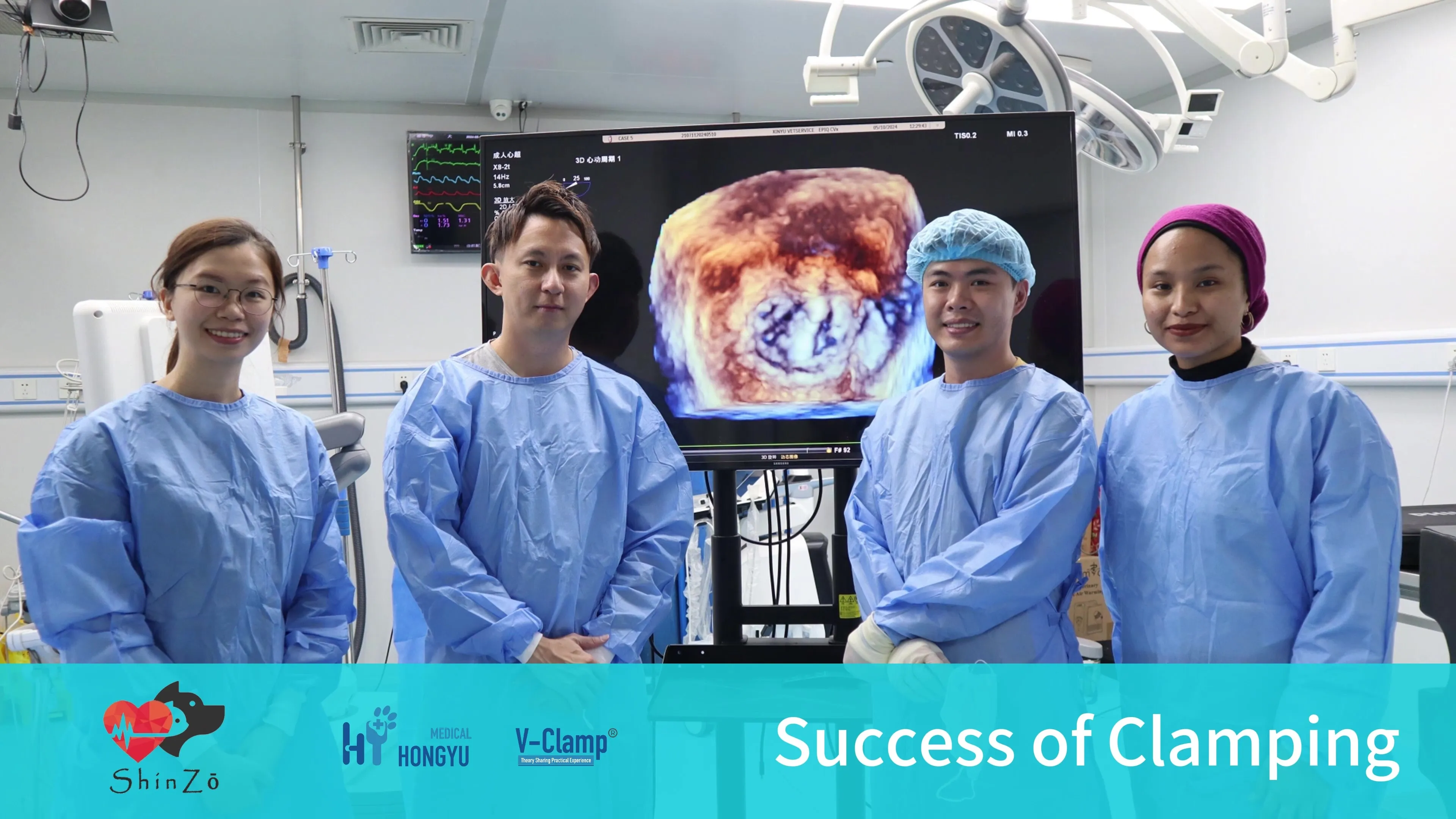
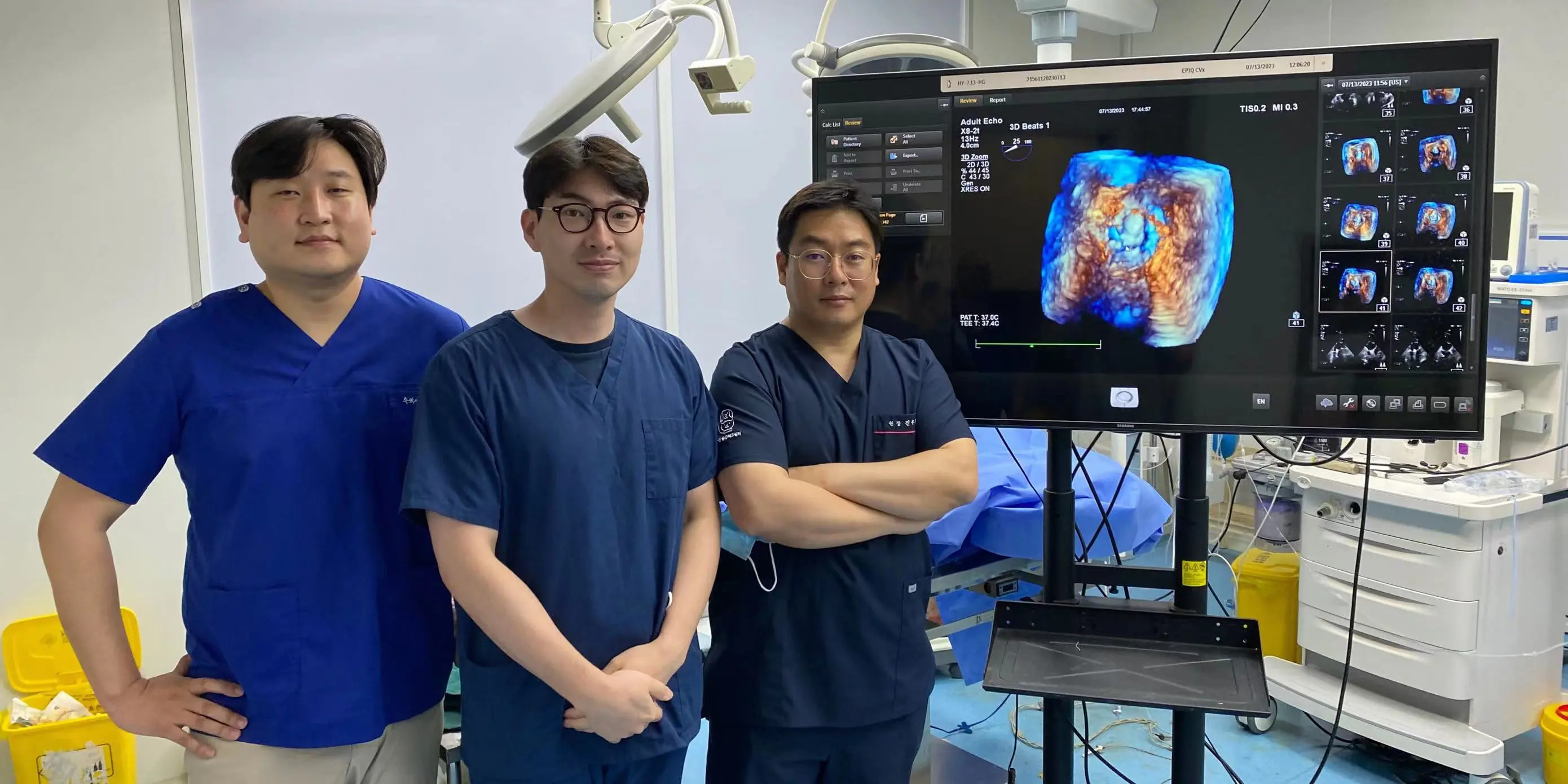
Recently, our Korean V-Clamp medical team has completed a series of highly challenging mitral valve repair surgeries on pet dogs, which is a record for both KAMC and Hongyu.
Case1
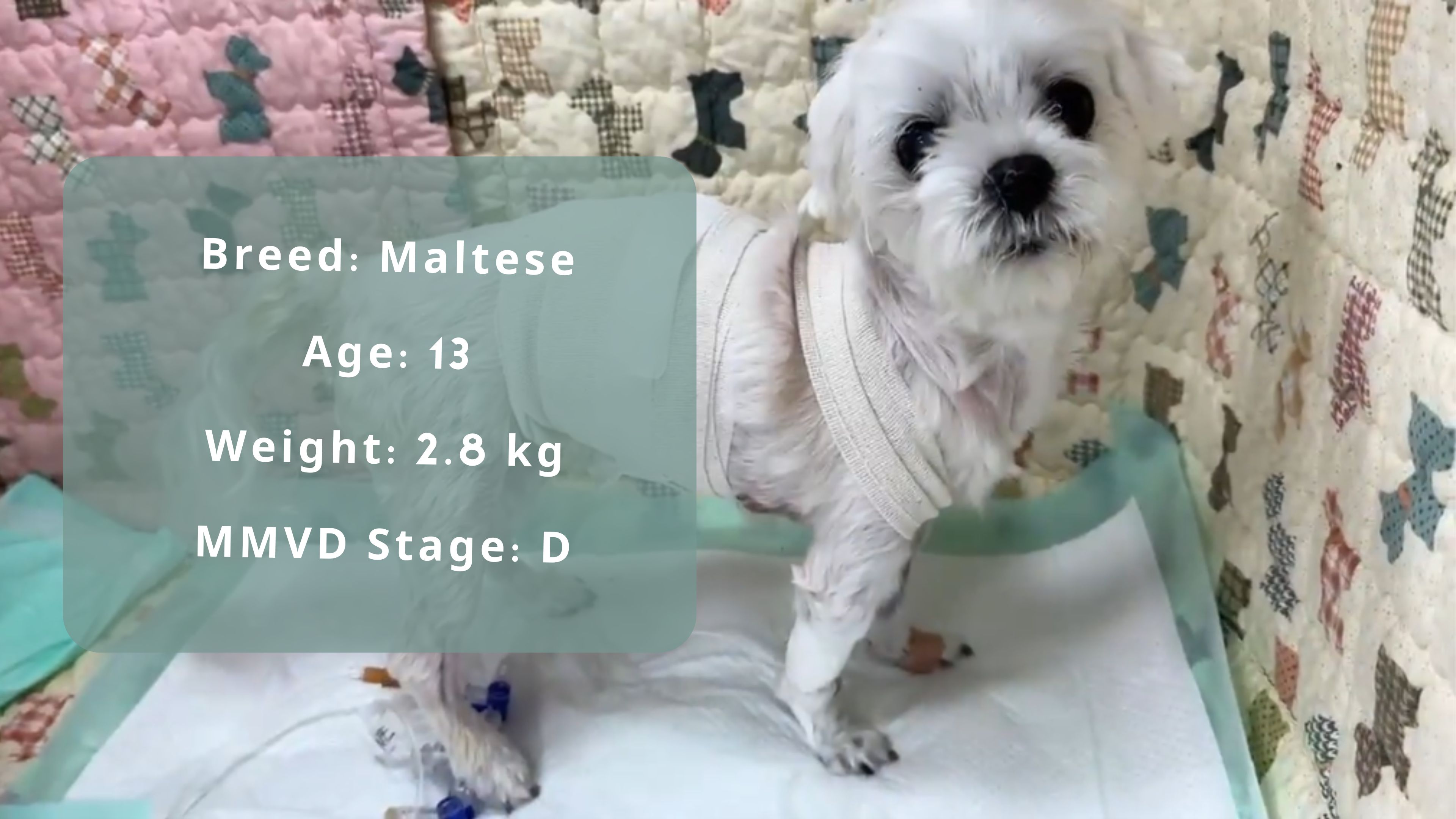
Yebbi has a very long history, diagnosed with pulmonary hypertension and MMVD Stage B1 in December 2020, and started taking sildenafil and pimobendan. By September 2021, the condition progressed to MMVD Stage B2, and an emergency visit for pulmonary edema in March 2022 led to a diagnosis of Stage C. By April 2024, the patient had visited the hospital nine times for pulmonary edema, with intervals shortening from nine months to three months, one month, two weeks, and finally ten days. Meanwhile, the patient was diagnosed with Cushing's syndrome and became blind due to SARD.
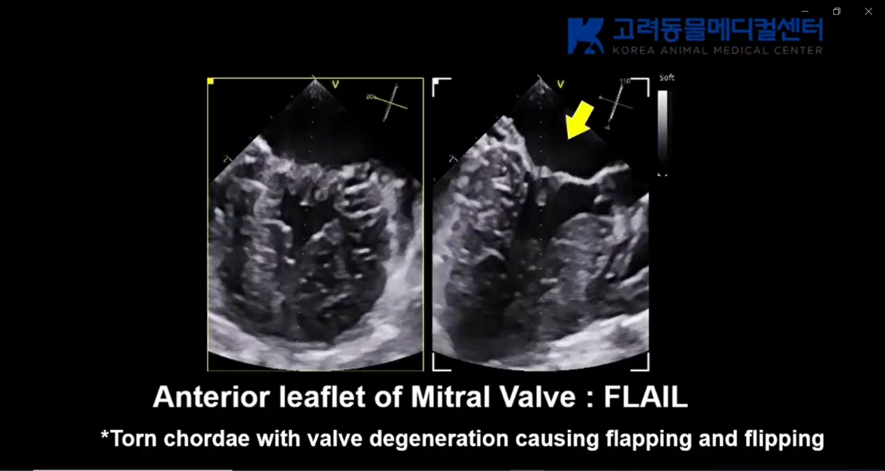
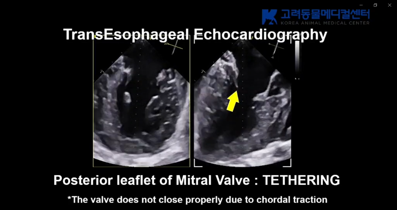
The patient showed flail of the anterior leaflet and tethering of the posterior leaflet on transesophageal ultrasound. There was also a small cleft or defect in zone 3 of the anterior leaflet, along with existing Cushing's disease, pulmonary hypertension, and systemic hypertension.
There is concern about how the condition will change after V-clamp surgery. If the V-clamp is perfectly positioned (i.e., if MR is reduced too much), pulmonary hypertension and systemic hypertension might worsen. The patient might require more anticoagulants due to a higher tendency for thrombosis caused by Cushing's disease, or conversely, there might be an increased tendency for bleeding.
Despite these worries, the patient doesn't seem to have much time left. There aren't many images of Stage D patients during surgery because saving them takes time, and it's essential to minimize the surgery time for Stage D patients. Unlike other patients, the surgery for this patient required careful positioning of the V-clamp to avoid breaking the chordae or tearing the posterior leaflet due to tethering.
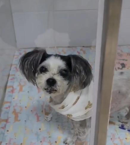
Post-surgery recovery, which was initially a concern due to the patient’s age and heart condition, went surprisingly well. Blood pressure during the V-clamp surgery was 110-130, and post-surgery, it was 120-130. The patient recovered well from anesthesia, was mentally stable, and was up and about just two hours after the V-clamp surgery. Pain management was effective, and the patient did not exhibit any pain, which is fortunate since significant pain in pulmonary hypertension patients can lead to non-cardiogenic pulmonary edema.
Highlights
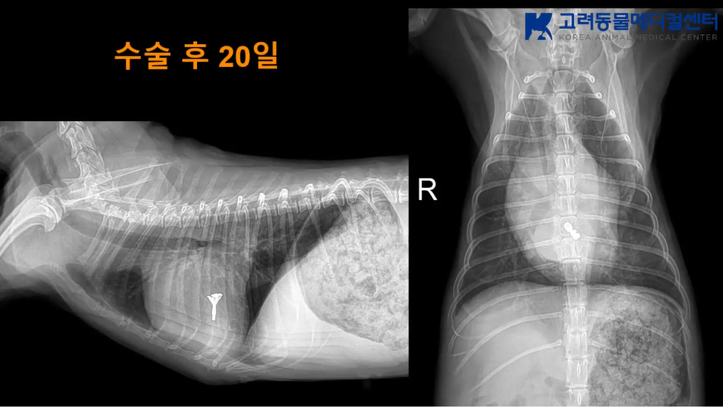
For Stage D patient, perfection should not be the goal; safety comes first. Despite this, Yebbi no longer has audible murmurs, and the radiography looks good. After discharge,Yebbi’s owner said she walks around, eats well, and sleeps well at home. The diuretic dosage was reduced to half of the original amount. Yebbi has gained some weight significantly, from 2.8 kg before the operation to 3.4 kg in short time. How much more it can be reduced will depend on Yebbi's pulmonary hypertension.
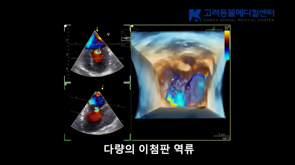
Pre-surgery (serious regurgitation)
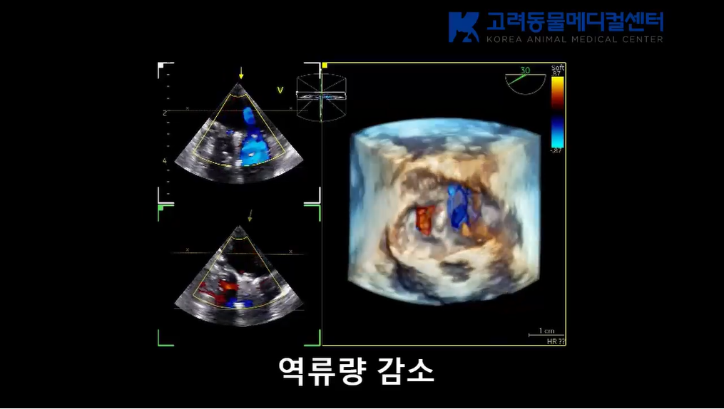
Post-surgery (much improved of regurgitation)
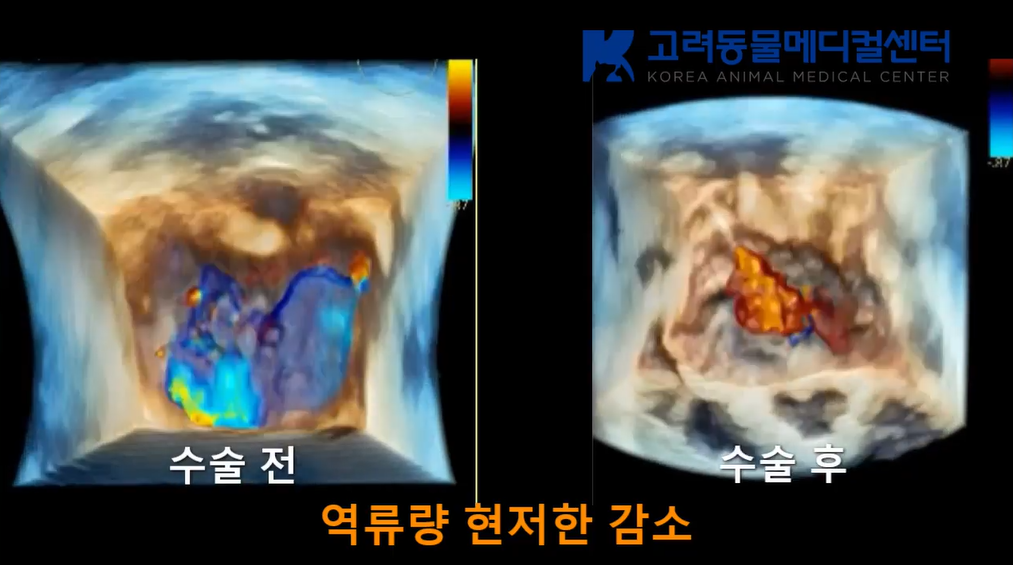
3D comparison before and after the surgery
Case 2
Details
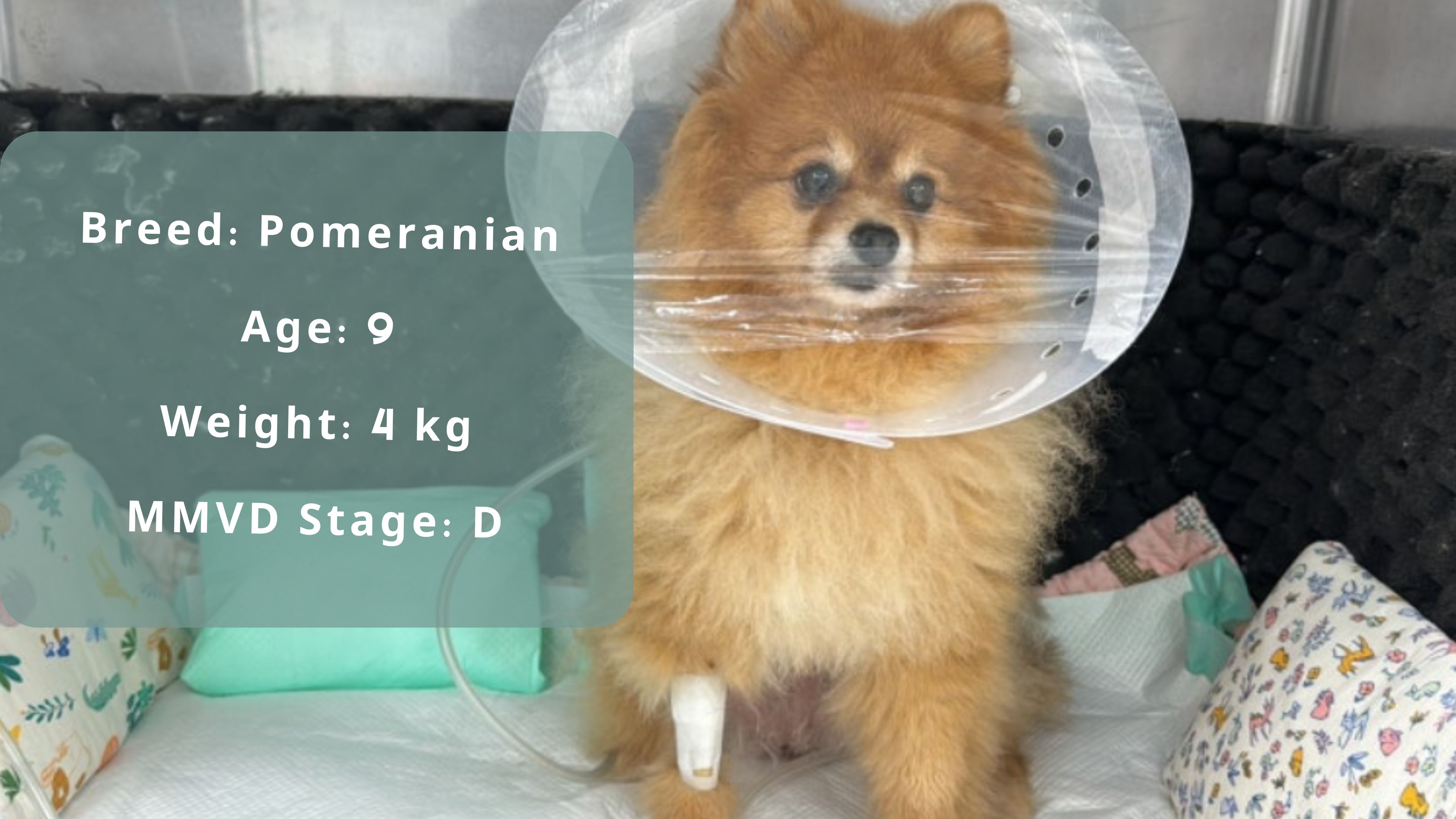
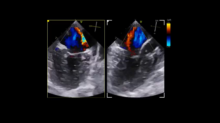
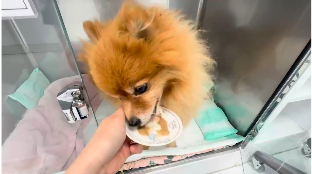
The 9-year-old Pomeranian patient visited a week ago for a health check-up and was found to have pulmonary edema on chest radio graphs. The patient had annual check-ups, but this year, due to poor condition and coughing, came in earlier than usual. The patient was hospitalized immediately for diuretic treatment, and after stabilizing the breathing, an echocardiogram revealed strong suspicion of bilateral chordae rupture (flail leaflets). The next day, a transesophageal echocardiogram confirmed severe regurgitation and flail in both the anterior and posterior leaflets. The primary veterinarian, unable to discharge the patient due to the risk of further chordae rupture, referred the patient for immediate surgery, especially given the distance of more than three hours from the clinic to the patient's home.
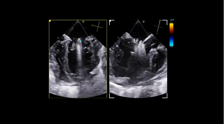
Just before surgery, a cleft or slightly short length in the posterior leaflet was suspected. The patient’s heart had a long and narrow shape, making it challenging to determine the catheter entry point. Due to the heart’s axis rotation, it was difficult to simultaneously ensure that both leaflets were properly captured between the clamps. In such cases, the procedure involved checking the anterior leaflet first, then the posterior leaflet, and then verifying in 3D, more frequently than with other patients. Additionally, since it was a flail leaflet, it was essential to close the clamp during diastole. As the clamp approached the valve, the heart rate increased, requiring the surgeon to synchronize their breathing pattern with the heart rate and close the clamp accordingly. Since both sides couldn't be seen at once, it took longer to measure and confirm adequate capture of the leaflets on still images before releasing the clamp, to avoid tearing the valve. This process felt very long and slow, filled with tension.
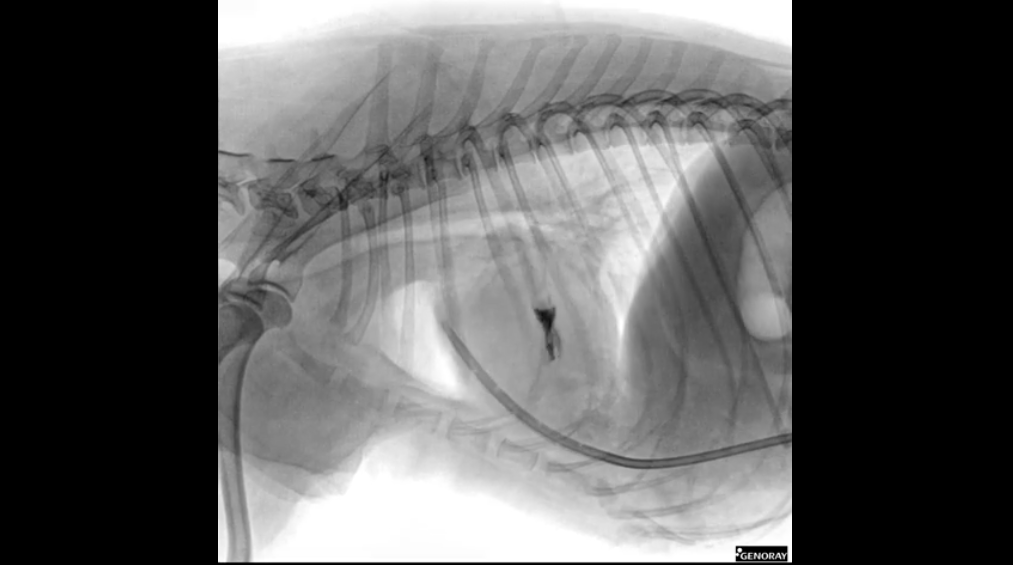
Fortunately, the clamp captured a sufficient amount of the leaflets, and the patient’s recovery was good. Mental recovery is crucial after V-Clamp surgery. The effective post-surgery pain management and return to normalcy of the patient’s mental state indicates a good prognosis. If the mental state does not improve, it might suggest a serious underlying issue, though the extent varies with the patient’s age and underlying conditions.
The next morning, the patient’s blood pressure, heart rate, body temperature, and respiratory rate (18–24 breaths per minute) were all normal. The first night after V-Clamp surgery is critical. Adequate pain management and stable vital signs are essential, with mental state and blood pressure being the most important.
Pre-Surgery
During the Surgery
Outcomes
Post-Surgery
Case 3
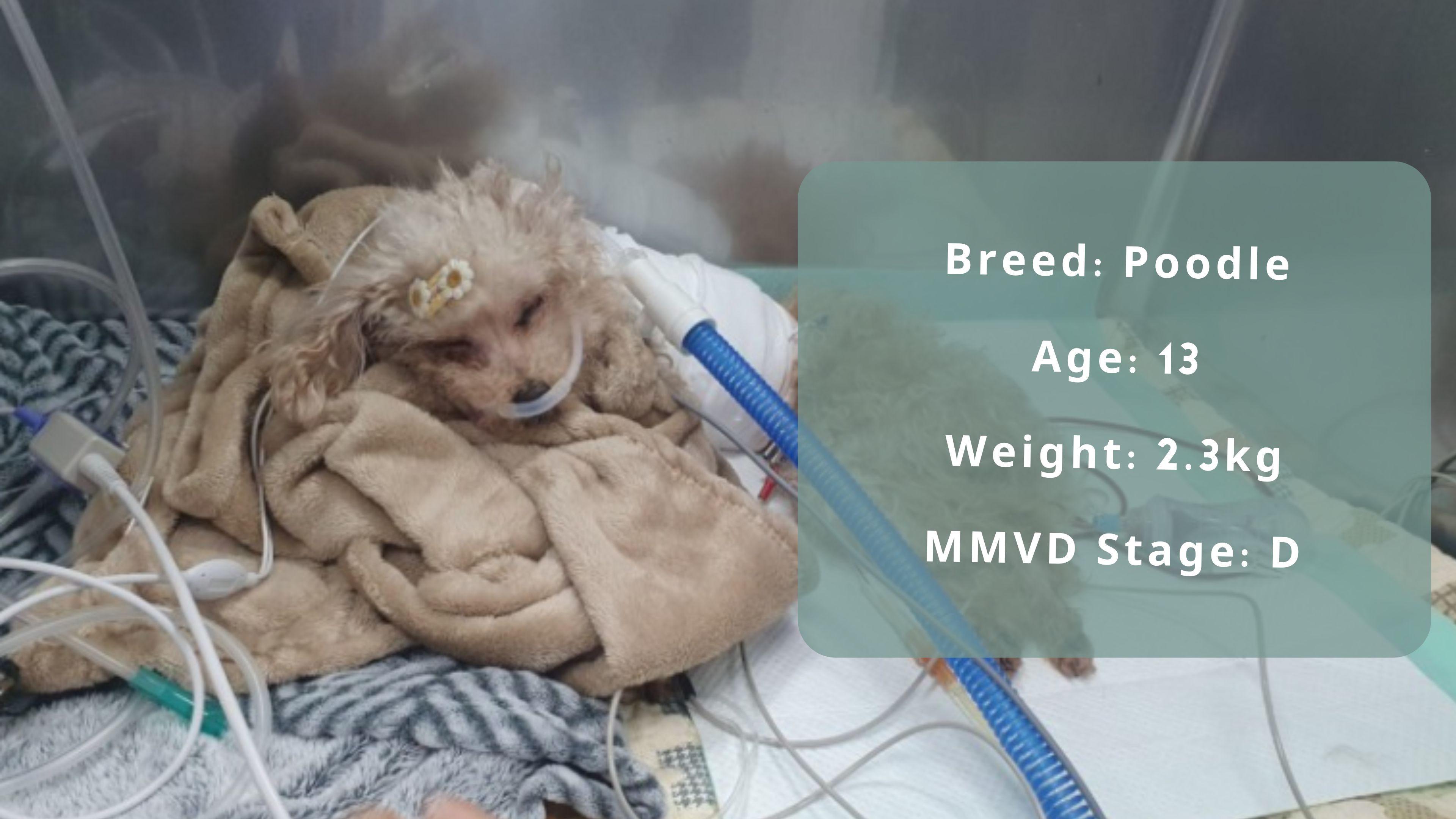
This patient was admitted with pulmonary edema. Initially presenting with a cough, there were no severe heart murmurs. However, three days later, the patient was urgently readmitted with severe heart murmurs, indicating a high possibility of chordae rupture due to the rapid progression.
The patient underwent a transesophageal echocardiogram, but the surgery decision was delayed by the guardian due to the high risk. Over the next month, the flail of the valve worsened, and the pulmonary edema did not respond well to high doses of diuretics and inotropes.
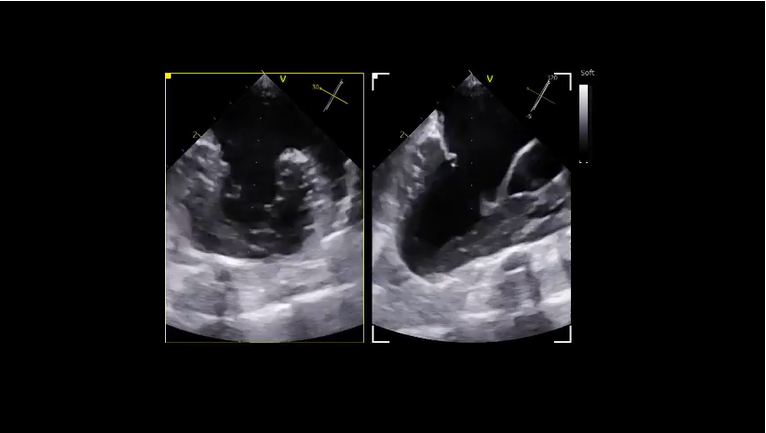
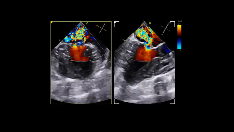
This patient was previously mentioned in the [V-Clamp Series] regarding tethering, where valve prolapse led to flail, which rapidly progressed to Stage D within a month. The patient weighed only 2.3kg, had a severely flail anterior leaflet, a short posterior leaflet, and preoperative MR (regurgitation) was 87%. The myocardial thickness had decreased, and myocardial function had significantly declined compared to a month ago.
Given the severely compromised heart function, there was a high risk of postoperative mortality. The patient required absolute rest immediately after surgery.
Discussion
Fortunately, the patient's blood pressure and heart rate stabilized overnight, allowing for a reduction in vasopressors. The next day's radiographs also looked promising. The V-clamp angle was good, and there was no pulmonary edema despite administering fluids at 5ml/kg/hr without diuretics. The cardiomegaly appeared slightly improved, and the pulmonary infiltrates in the right caudal lobe seemed to be due to positional atelectasis.
There is still a long road ahead, but fortunately, the direction is toward recovery. The patient, after removing the chest tube and ECG monitor, got up to drink water. The most crucial criterion for predicting the prognosis after V-Clamp surgery is the mental state. The patient has regained consciousness and can stand up, which is a very positive sign.
Pre-Surgery
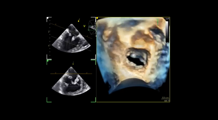
During the Surgery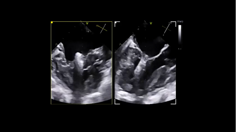
Post-Surgery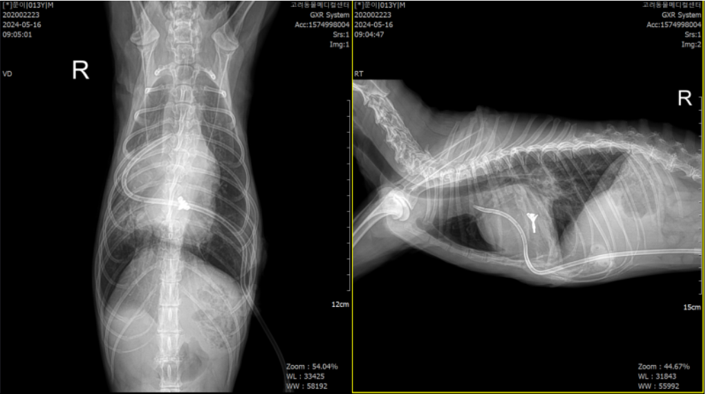
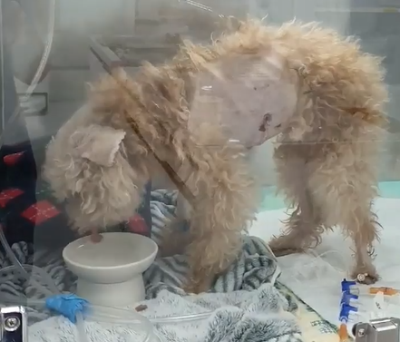
Case 4
Closing Notes
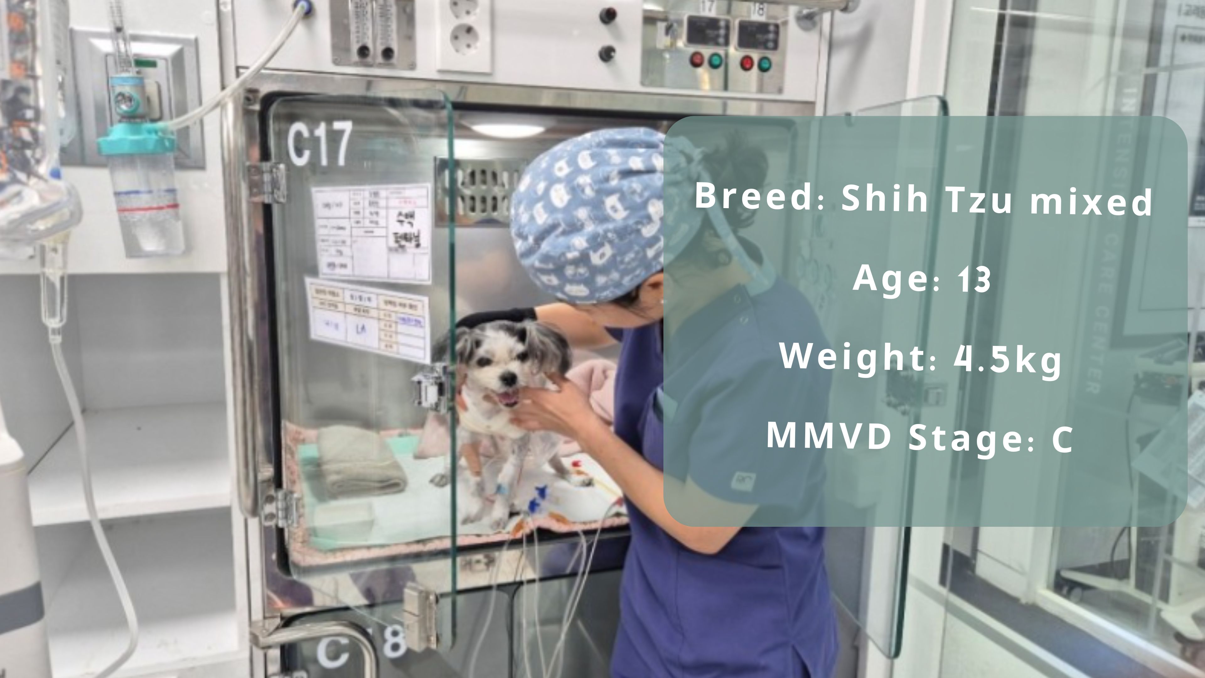
Daepal recently visited the emergency room for pulmonary edema. Daepal’s valve condition was very similar to Jaju's (a previous patient who happens to be Daepal’s sibling), they both had flail of the anterior leaflet and a cleft in the posterior leaflet. Such cases involve risks: if there's a cleft in the valve, it can tear during surgery or a few days afterwards.
Fortunately, the surgery proceeded without major issues. Daepal had pectus excavatum (a chest deformity), making it very challenging to establish the access point, which affects the quality of intraoperative imaging and the difficulty of the surgery. Two hours after the surgery, it was remarkable to see the 13-year-old patient up. And changes on POD (postoperative days) 1, 2, and 3 showed the heart undergoing reverse remodeling. This was the second V-Clamp surgery for a dog in this family with a genetic predisposition. Given Jaju’s good outcome, the guardian likely had high expectations, which added pressure to the surgical team. It's truly fortunate to see such positive results.
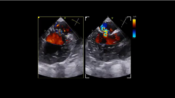
Pre-Surgery
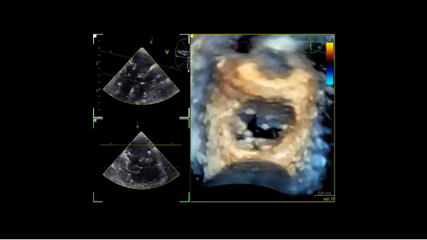
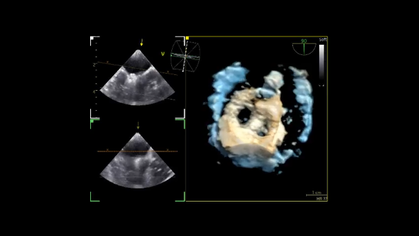
Post-Clamping
Statement of Source: Reposted from Doctor Youp's Blog