Case Background
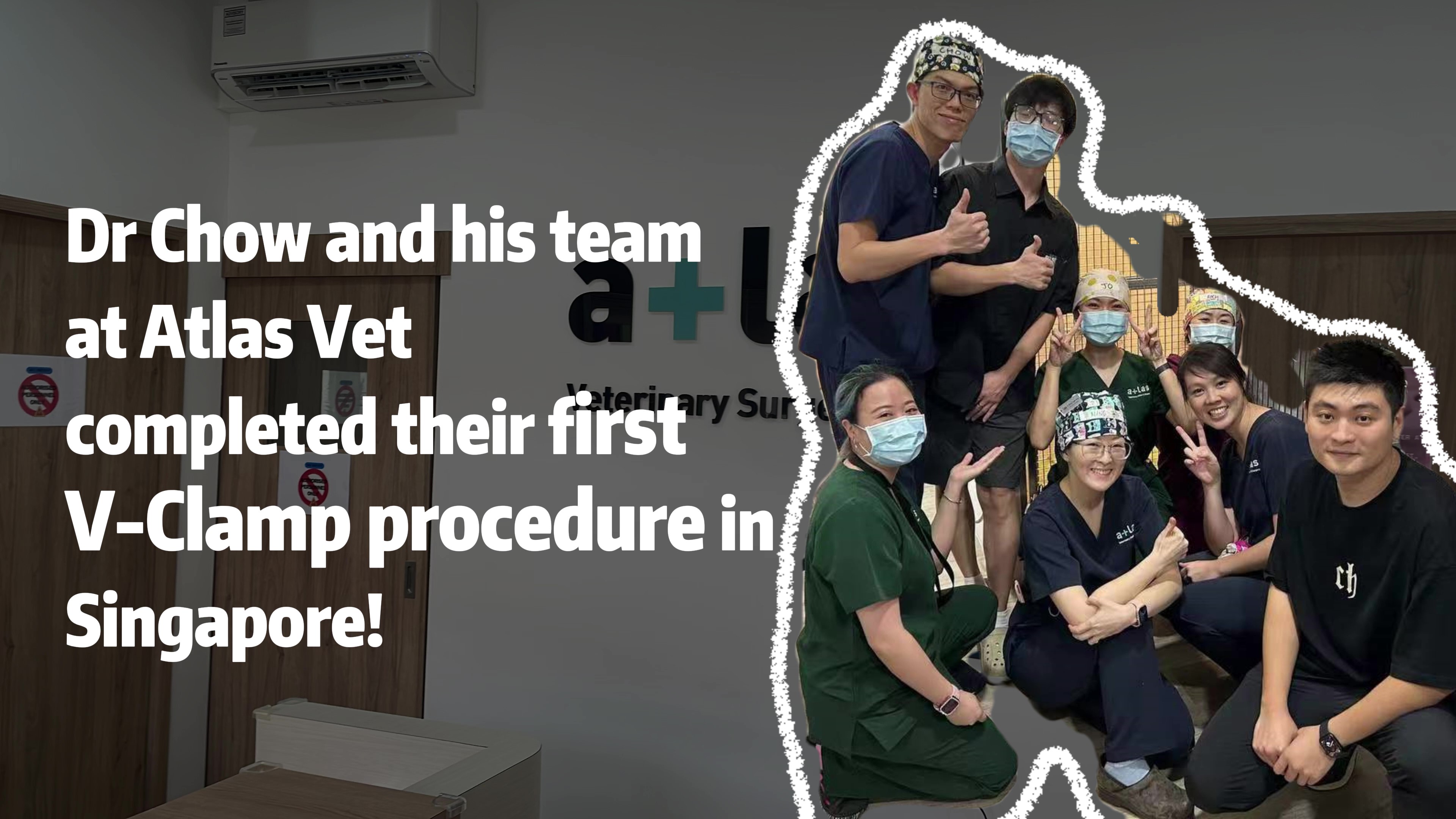
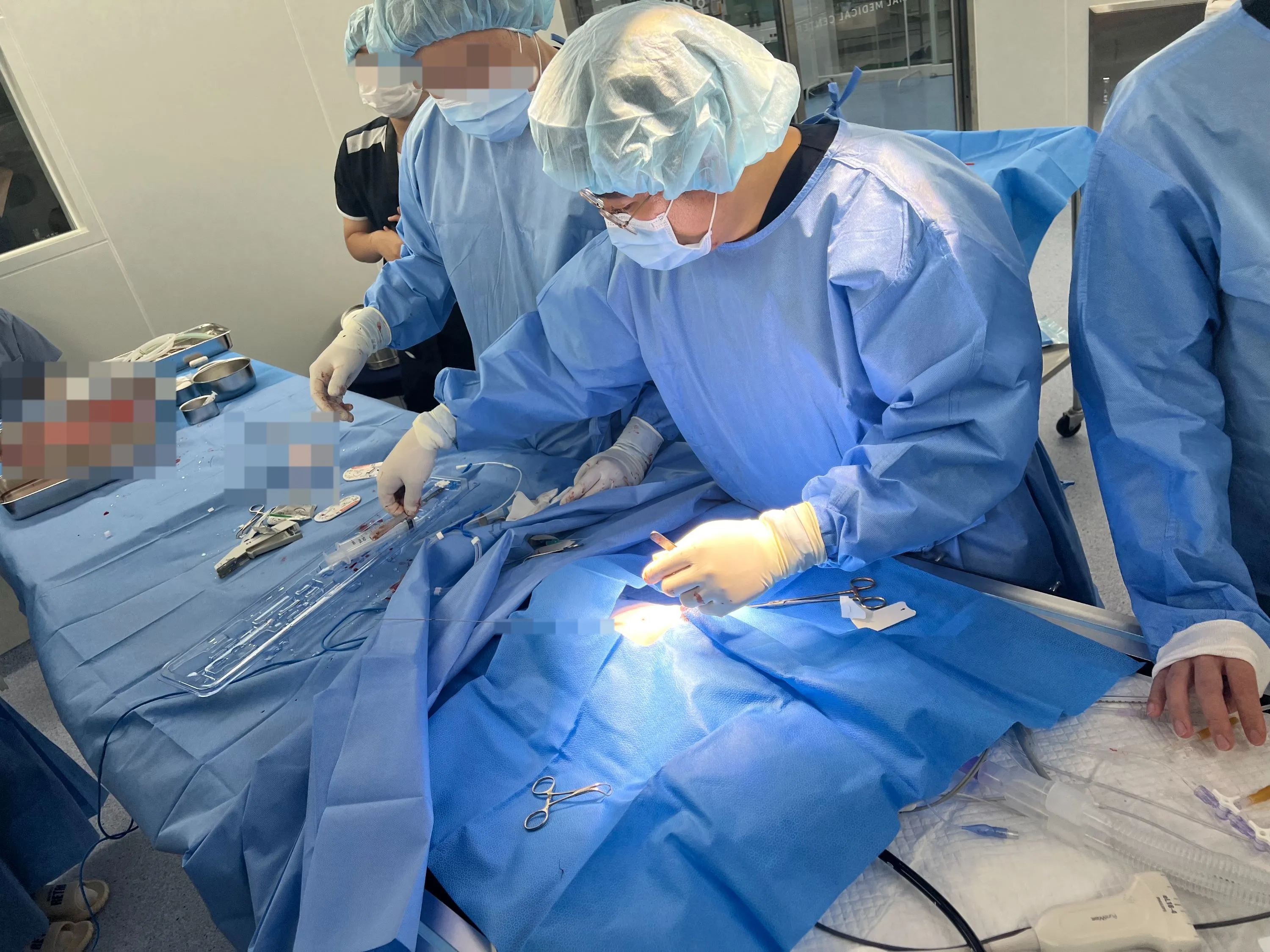
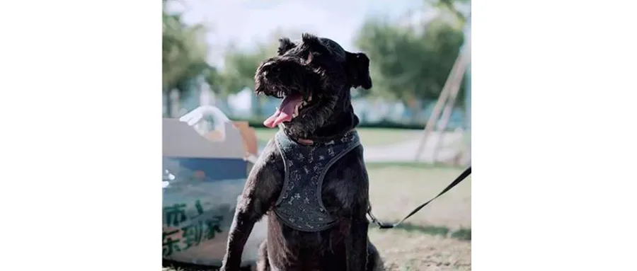
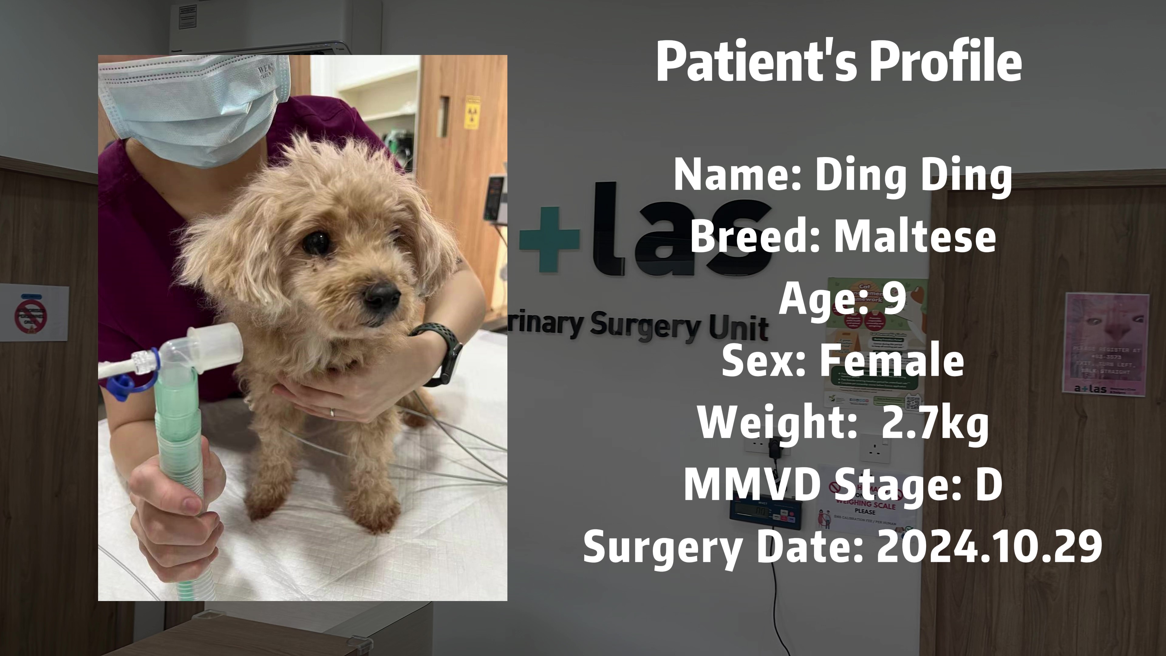
On October 29, 2024, Dr. Chow and his team at Atlas Veterinary Clinic & Surgery in Singapore successfully performed their First V-Clamp mitral valve repair surgery on DingDing, a 9-year-old female Maltese.
Clinical Presentation
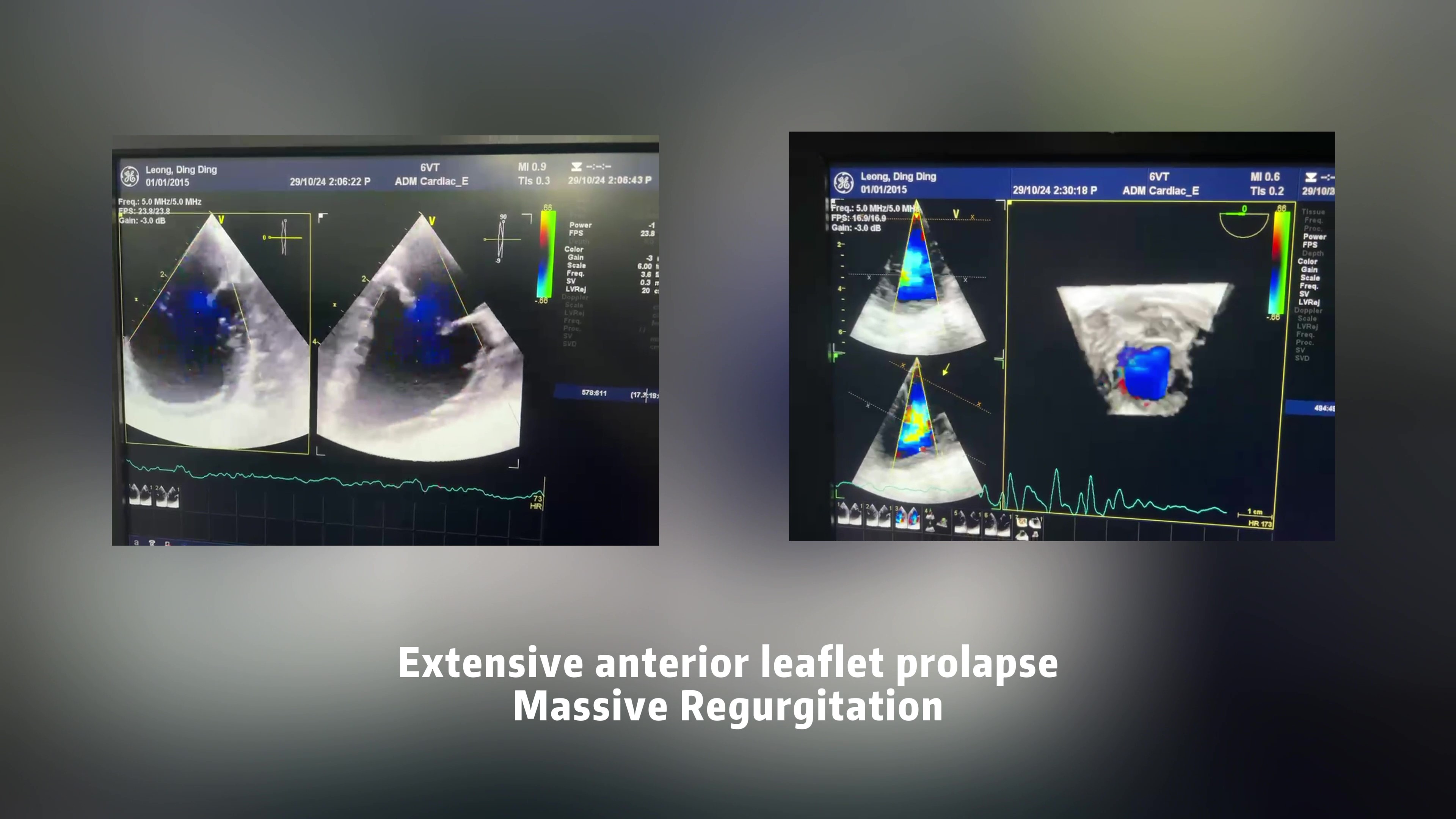
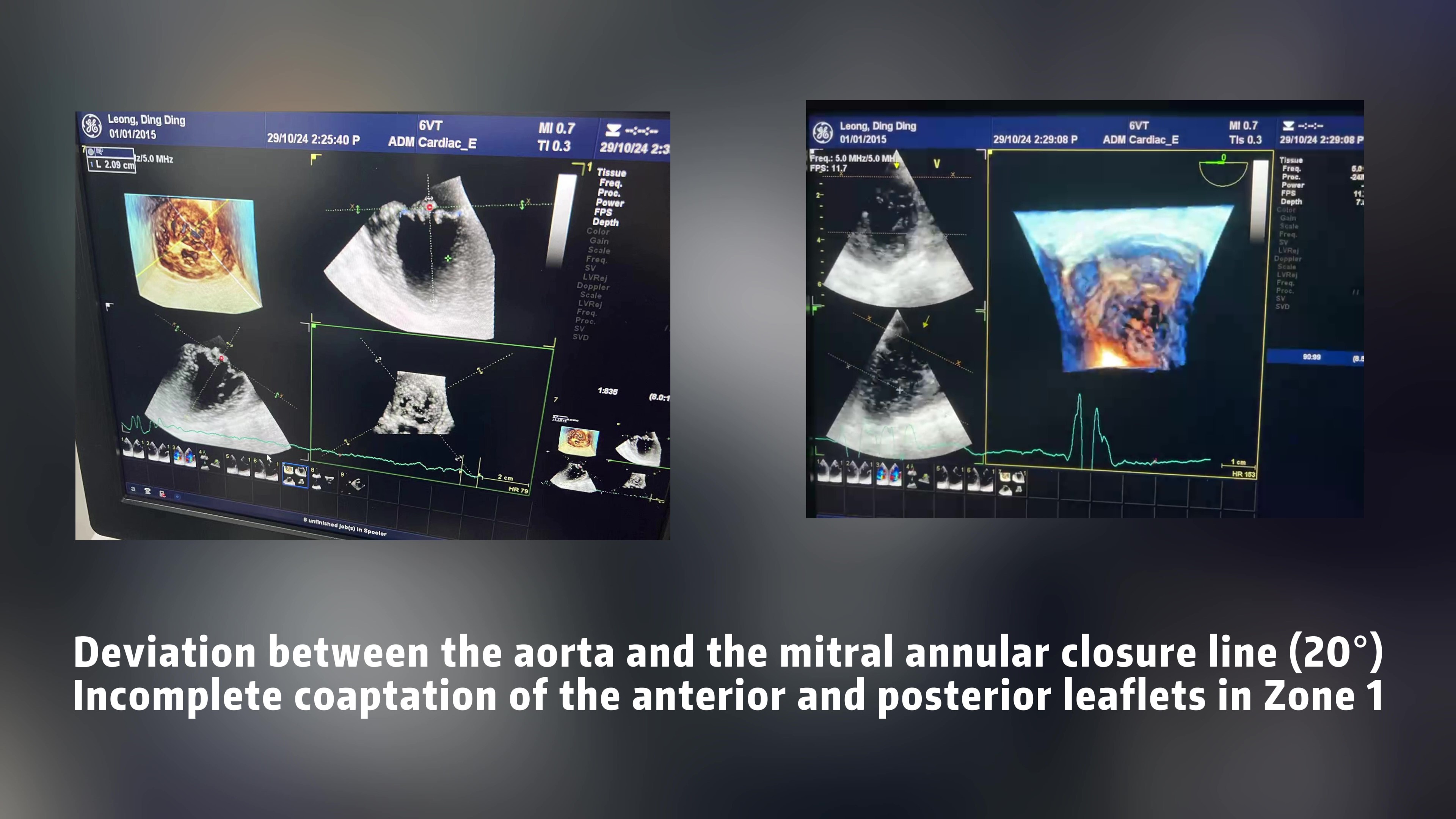
DingDing had been diagnosed with MMVD for over six months and was on medication to manage the condition. By the time of surgery, she had progressed to MMVD Stage D, which posed higher surgical risks. Preoperative transesophageal echocardiography revealed a mitral valve AP diameter of 18.5-20mm, with an anterior leaflet measuring 10.8mm and a posterior leaflet measuring 9mm. 3D imaging showed a 20-degree deviation between the aorta and the mitral valve annular coaptation line, with leaflet malcoaptation in Zone 1.
Preoperative
Procedure & Outcome
 Postoperative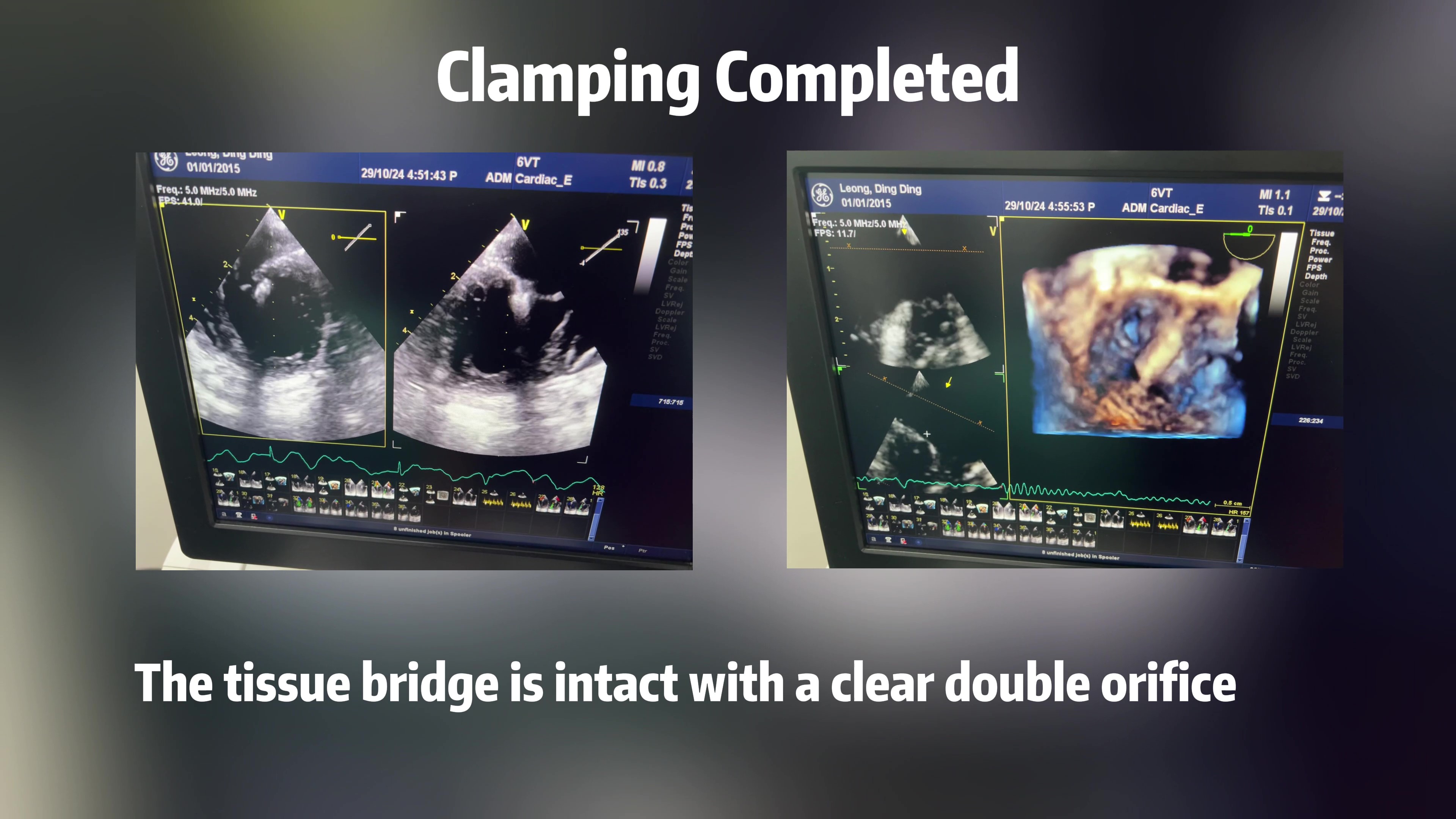
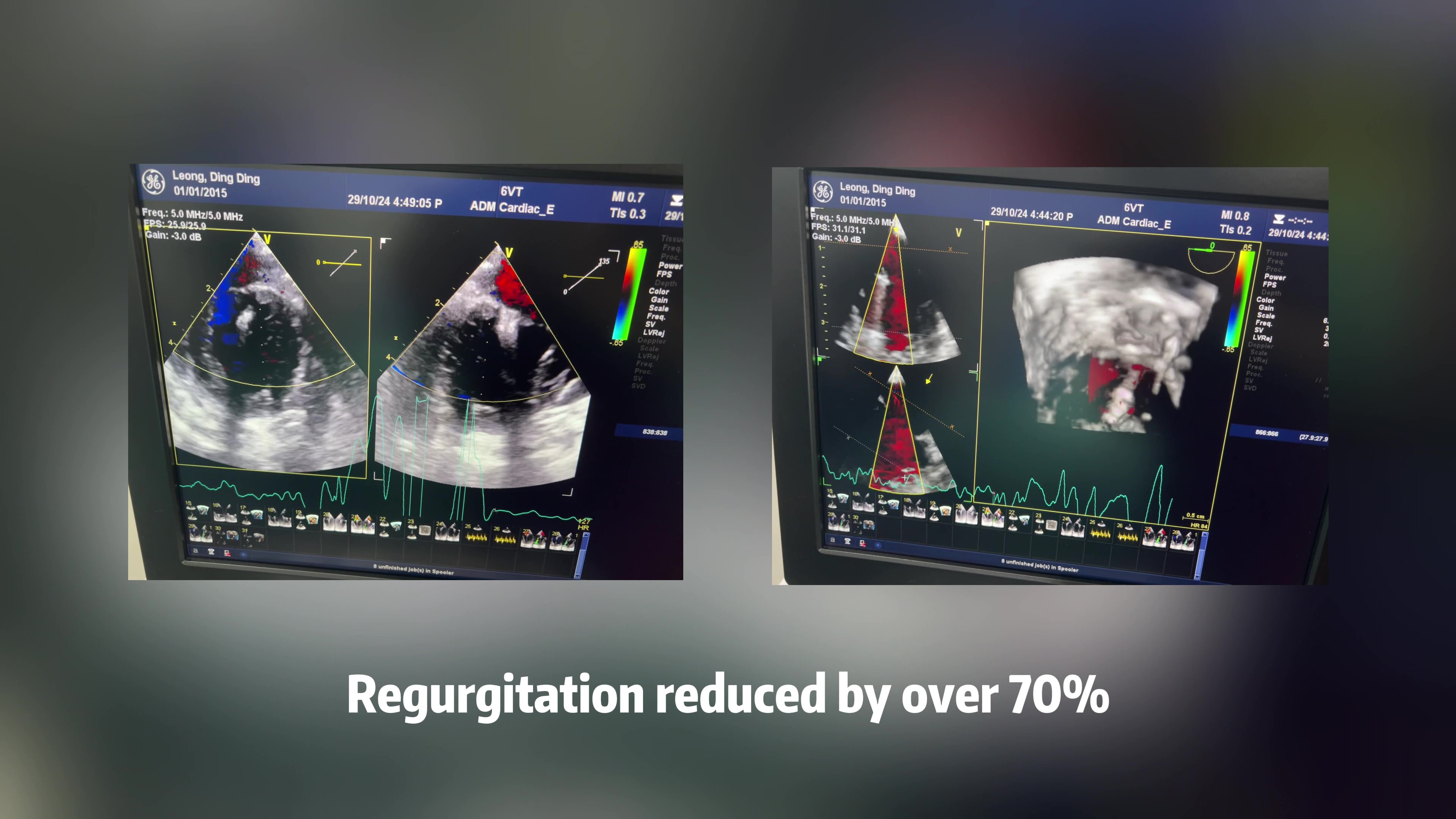
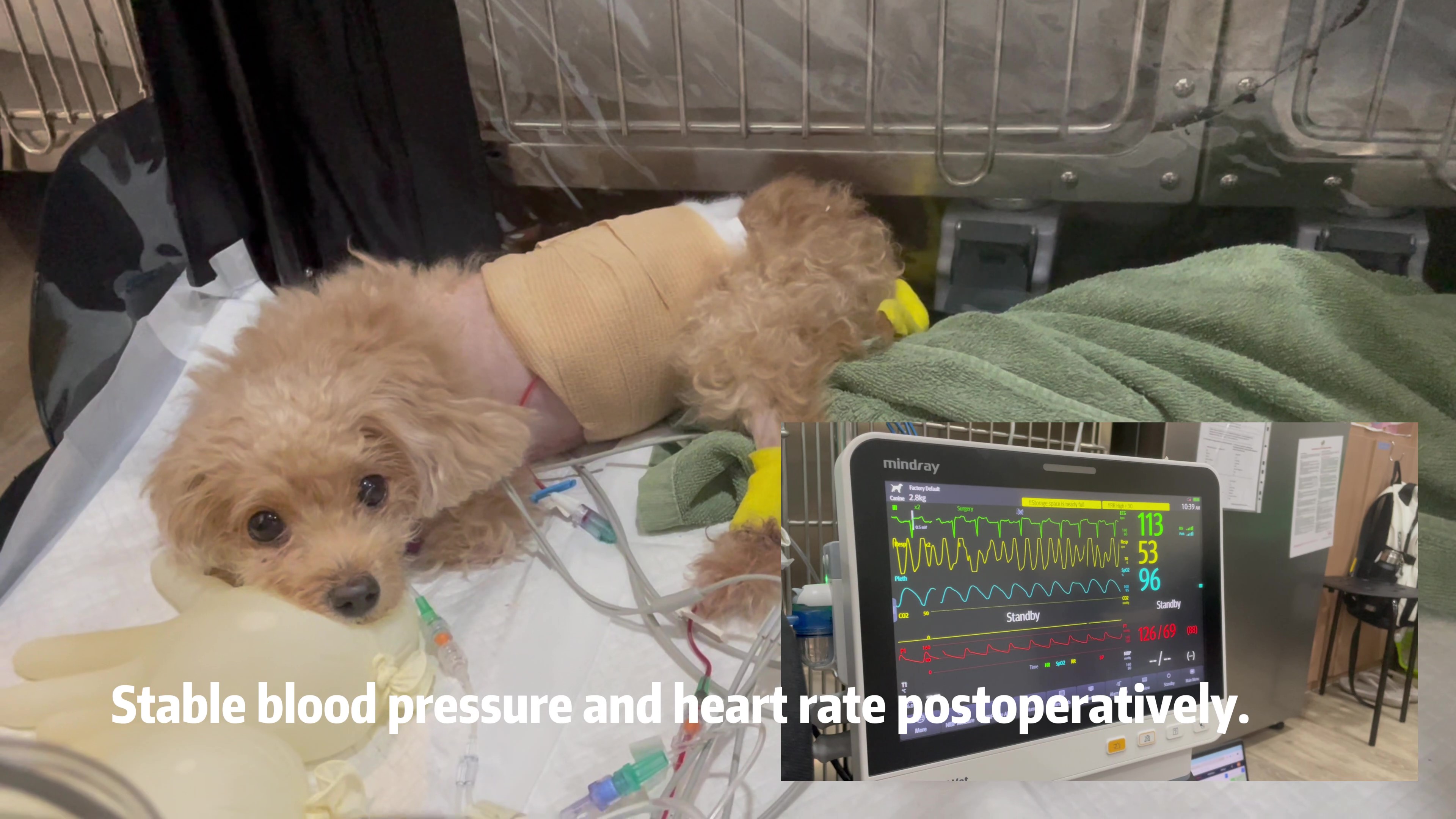
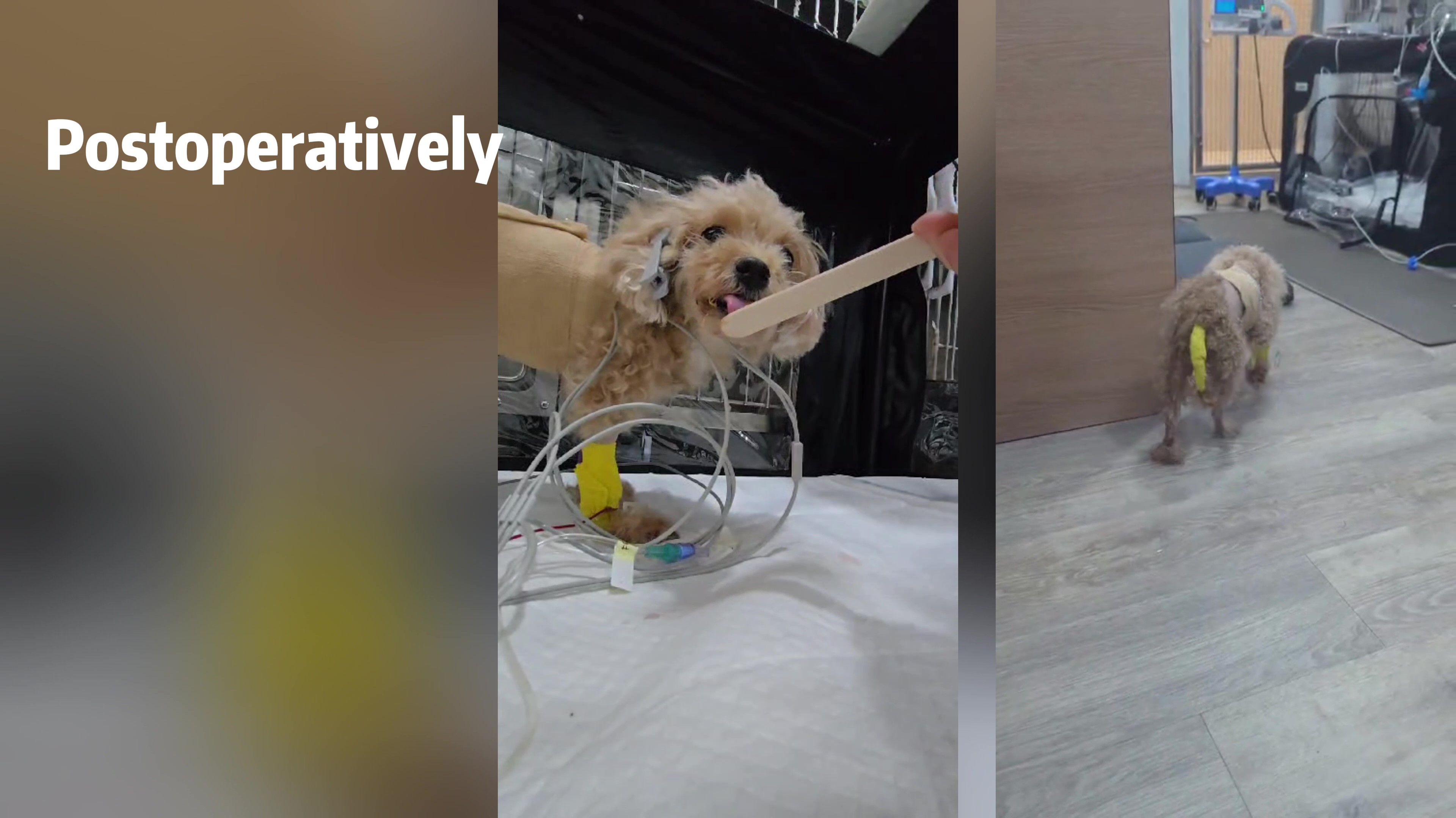
The surgery was successfully completed using a type Ⅳ V-Clamp applied to the A2P2 region. After clamping, regurgitation was reduced by more than 70%, with a small amount of residual regurgitation in Zone 3 due to the wide prolapse of the anterior leaflet. Postoperative 2D echocardiography confirmed stable clip placement, with an anterior leaflet grasp of 6mm and a posterior leaflet grasp of 7mm. 3D echocardiography showed the clip orientation as expected, at the 1 o’clock to 7 o’clock position, with an intact tissue bridge and clearly defined dual orifices. The mean postoperative transvalvular pressure gradient was 0.63mmHg, with stable blood pressure and heart rate.
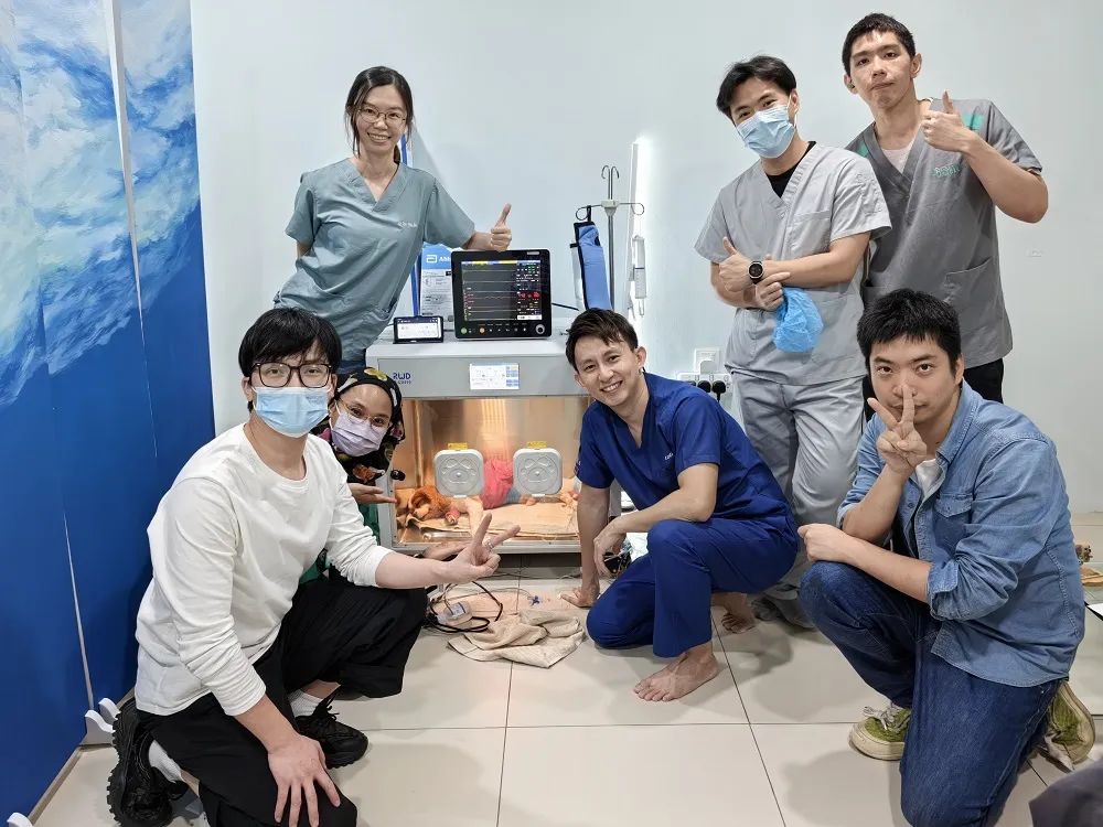
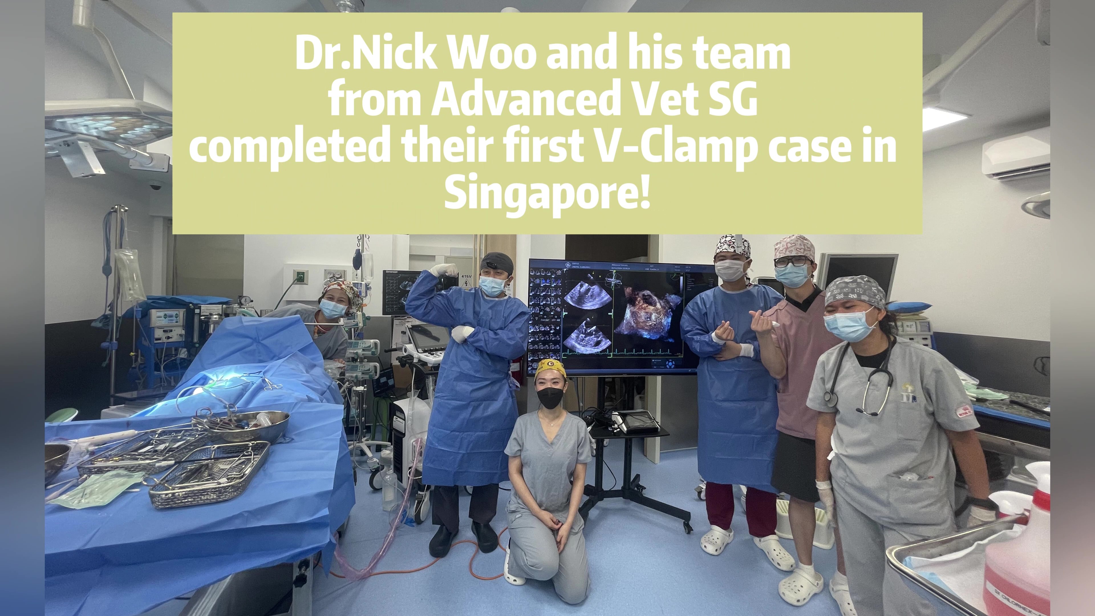 On November 1, 2024, Dr. Woo and his team at Advanced VetCare in Singapore successfully performed a V-Clamp mitral valve repair surgery on Toffee, a 9-year-old female Cavalier King Charles Spaniel diagnosed with MMVD.
Follow-up & Insights
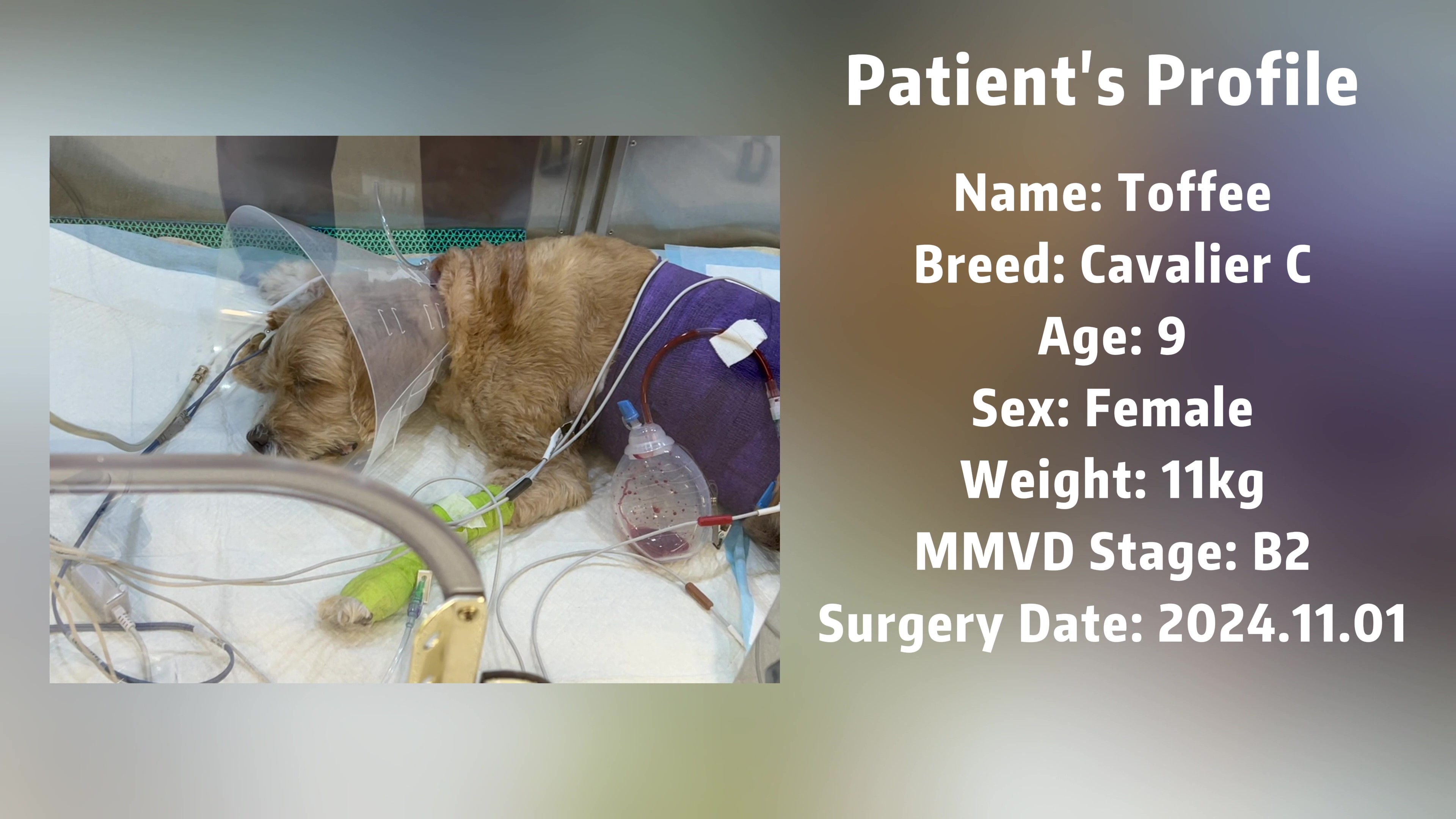
Prior to surgery, Toffee was in MMVD Stage B2 (B2 and early Stage C are considered optimal stages for surgery). Preoperative transesophageal echocardiography revealed a mitral valve AP diameter of 17mm, with an anterior leaflet measuring 11-12mm and a posterior leaflet measuring 7-8mm, both exhibiting prolapse.
Discussion
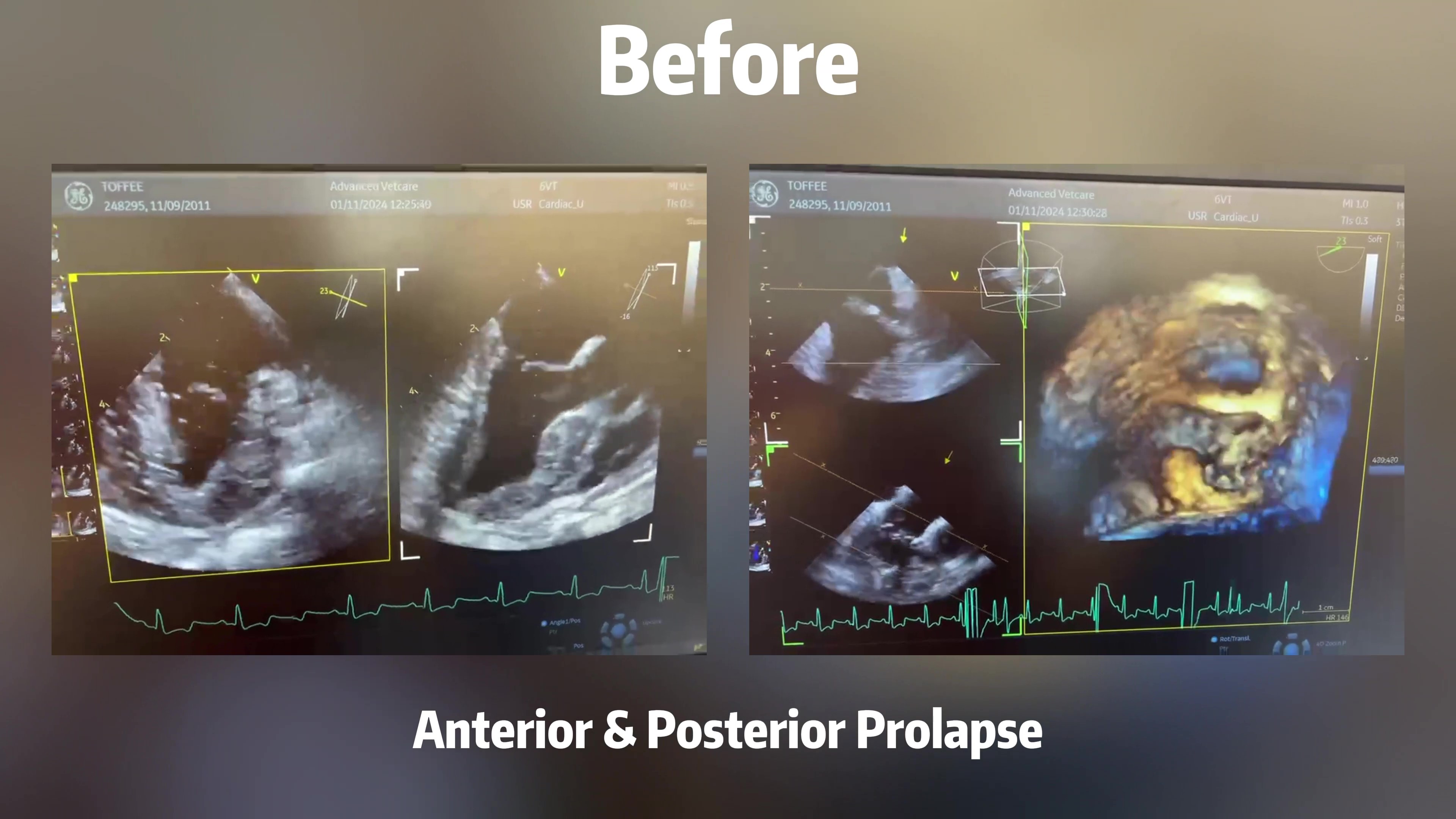
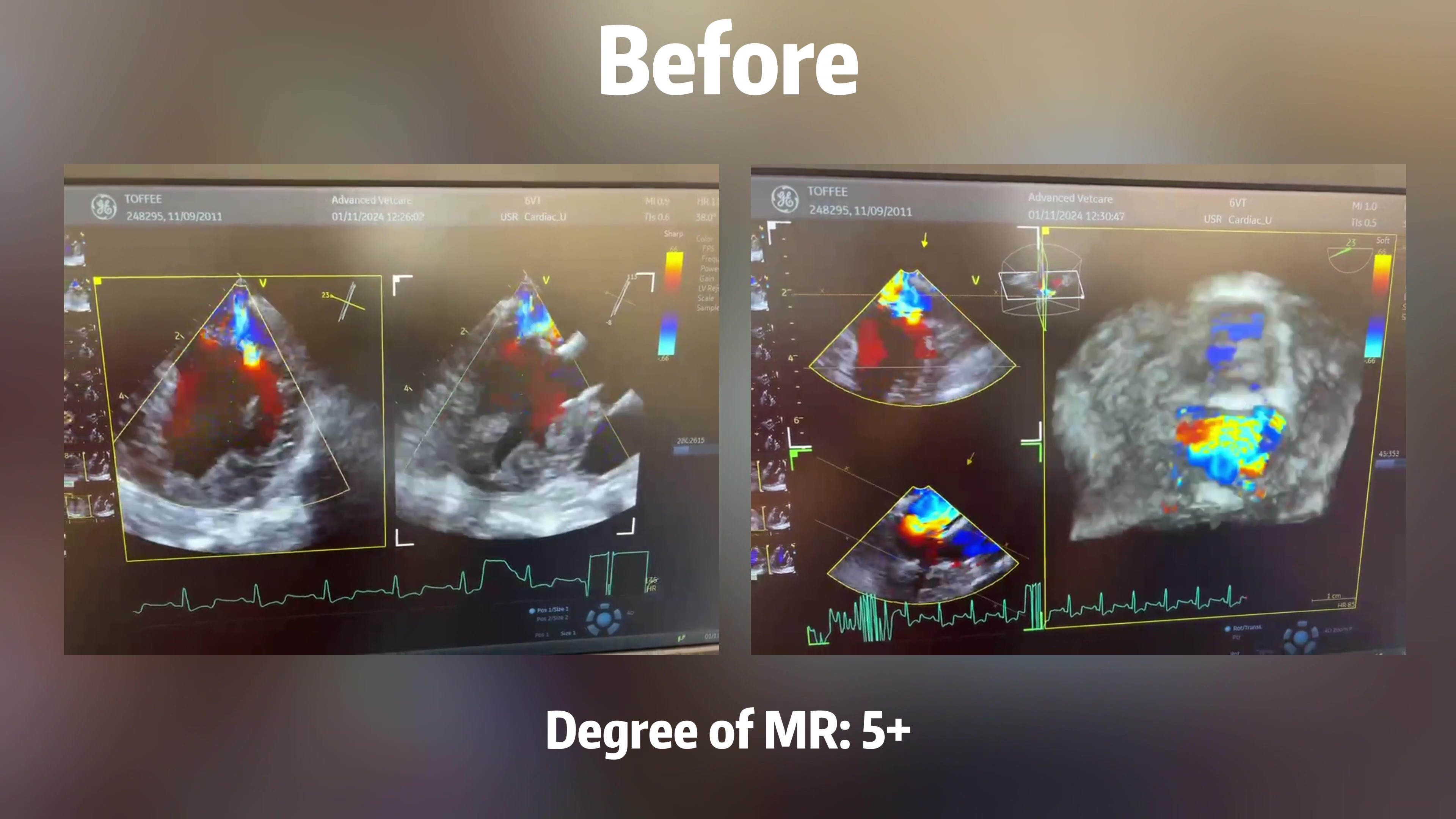
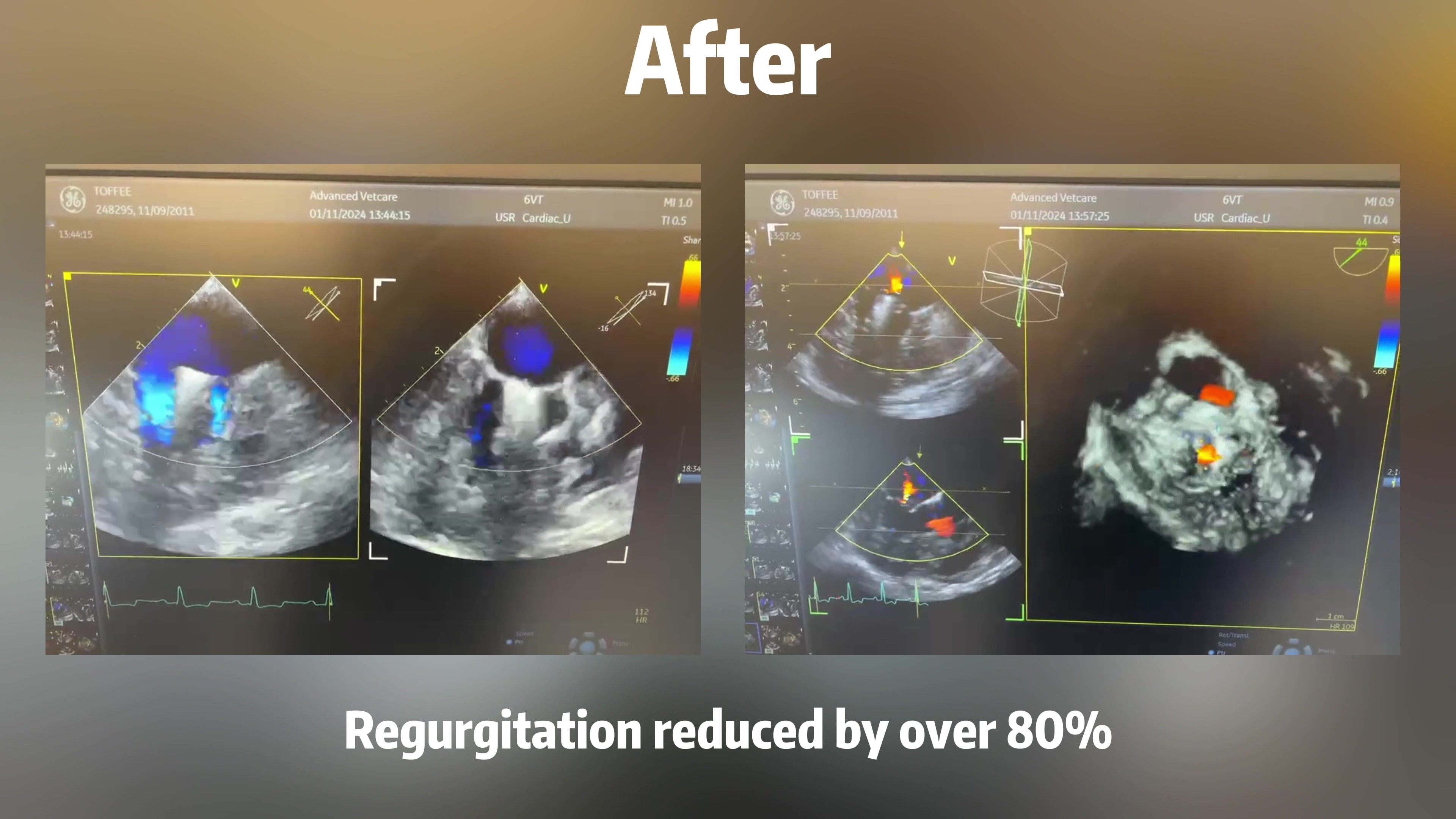
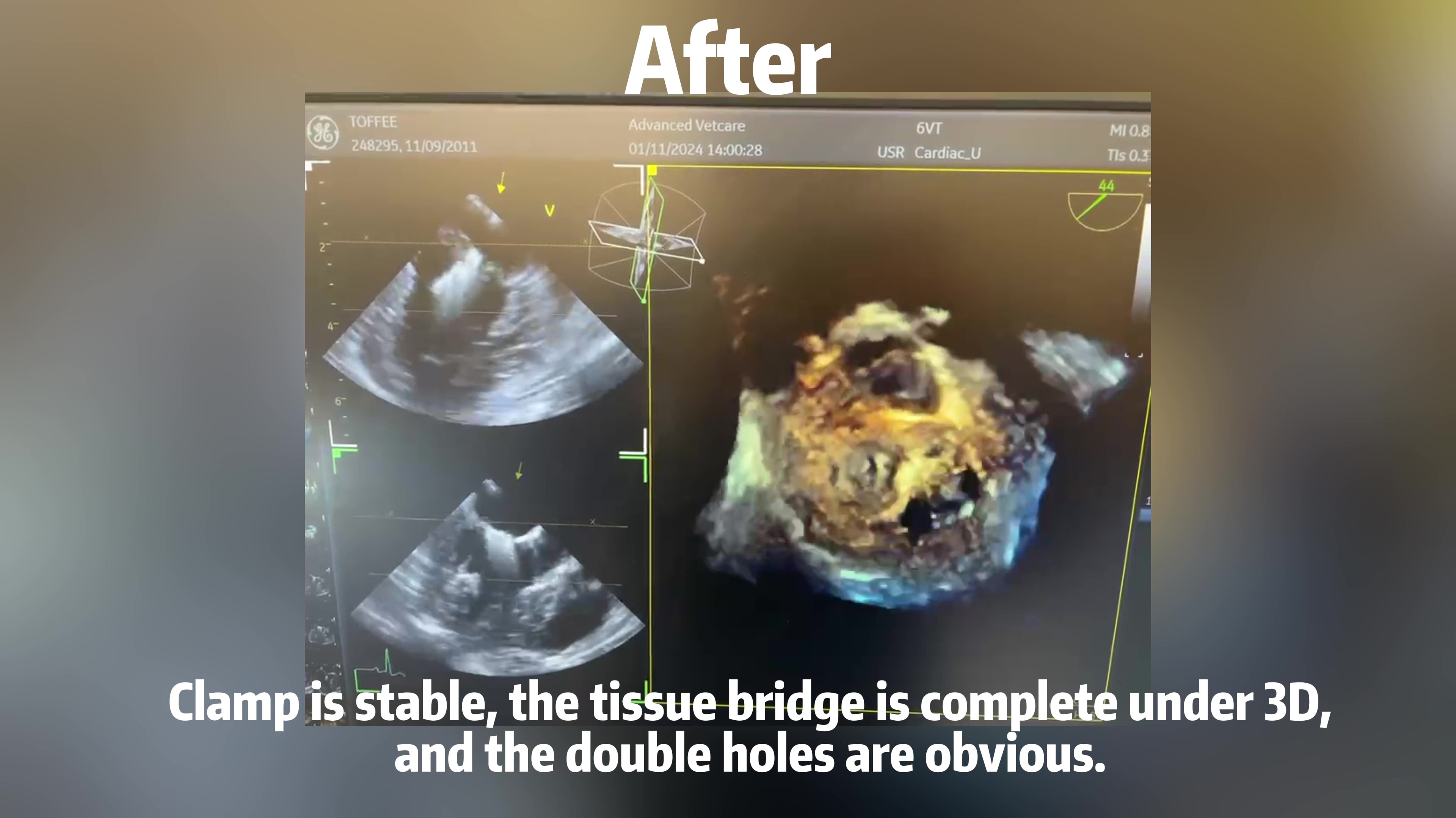
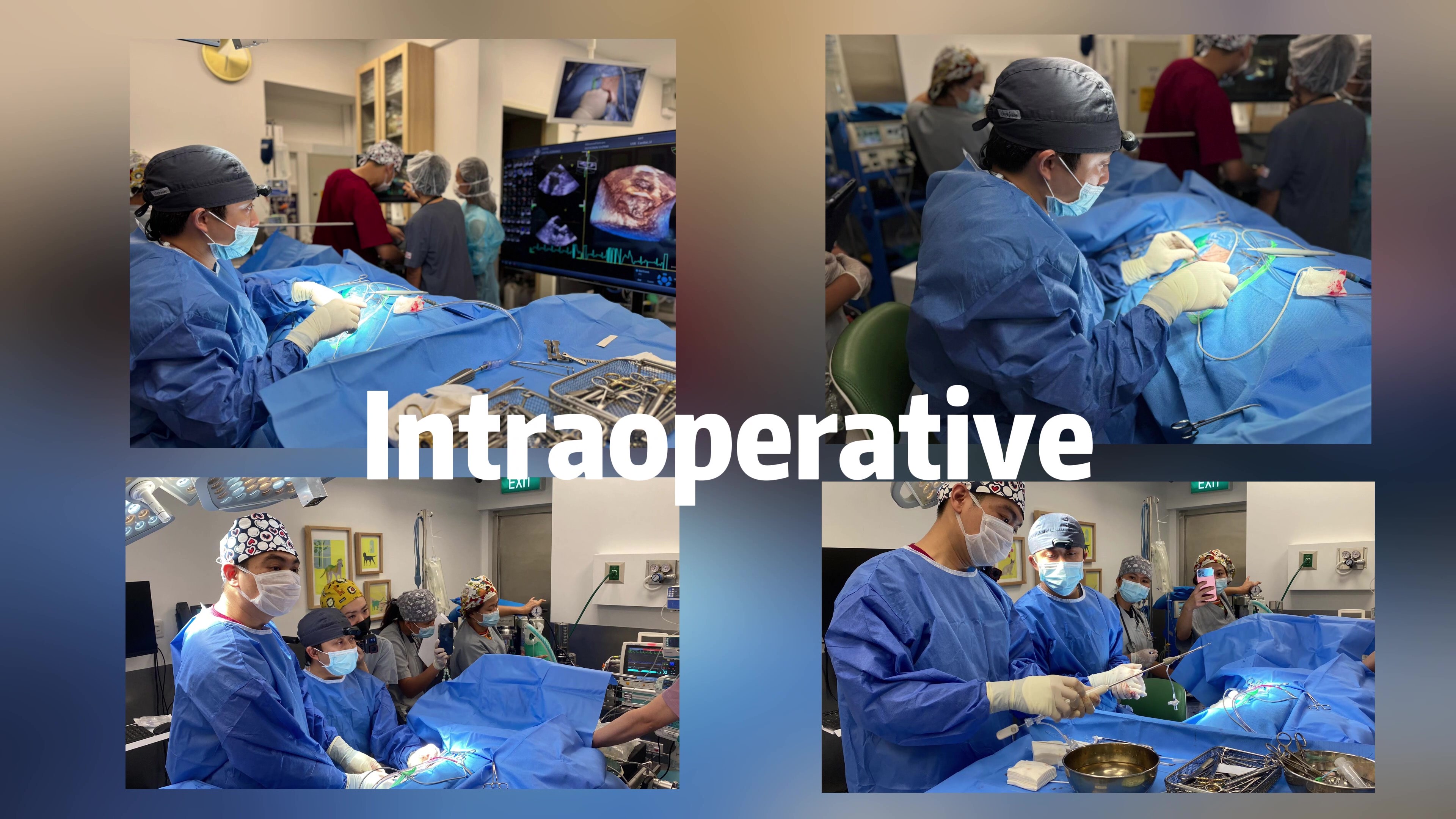
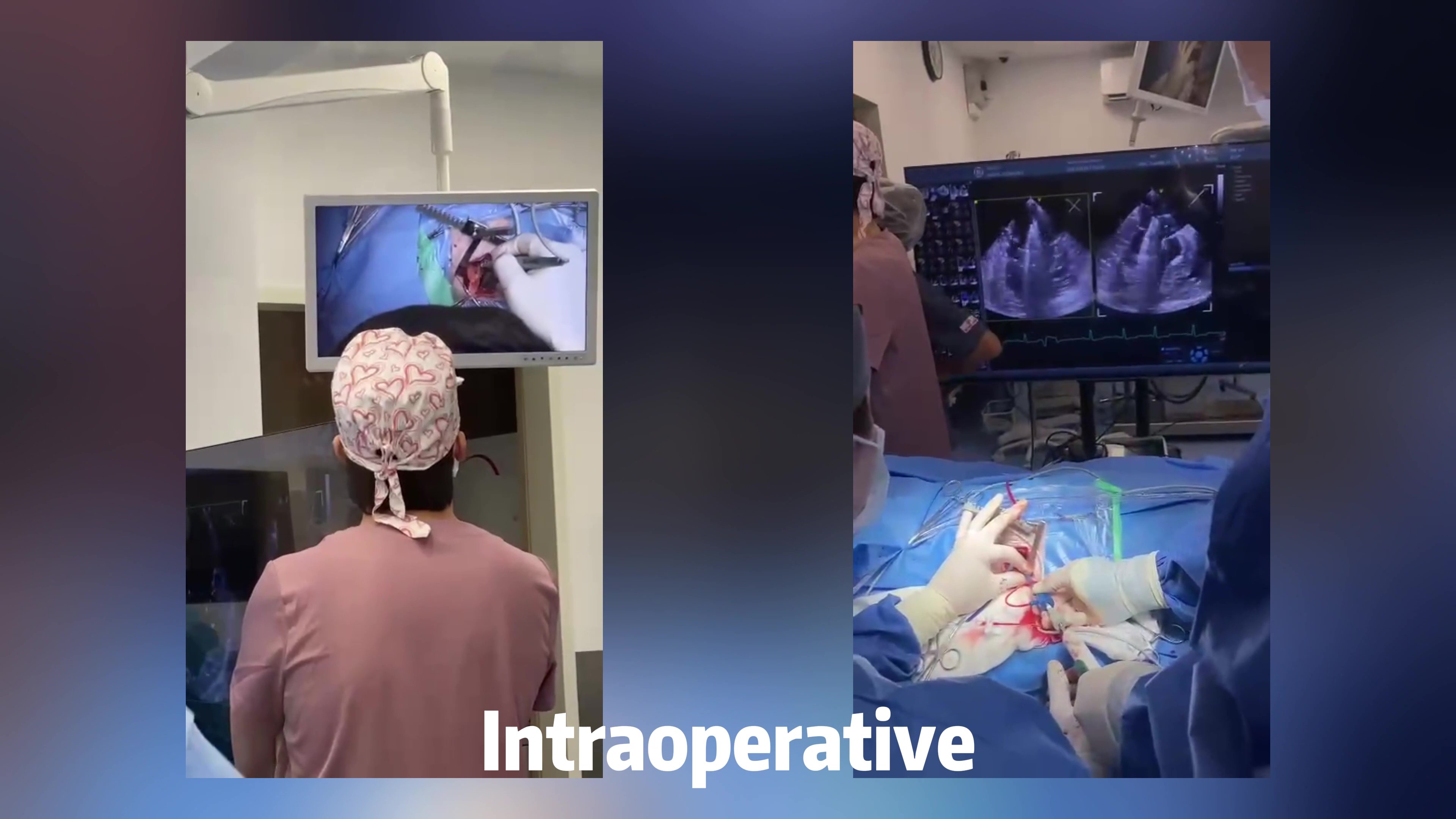
The surgery was successfully completed using a Type Ⅲ V-Clamp, applied at the A2P2 region, reducing regurgitation by more than 80%. Due to the angle deviation between the aorta and the mitral valve annular coaptation line, the clip was slightly oriented toward the 1 o’clock and 7 o’clock positions. Postoperative 2D echocardiography confirmed stable clip placement, with an anterior leaflet grasp of 8mm and a posterior leaflet grasp of 5mm. 3D echocardiography showed an intact tissue bridge and clearly defined dual orifices. The mean postoperative transvalvular pressure gradient was only 0.8mmHg.
Closing Notes
The successful completion of the two V-Clamp procedures marks a significant milestone, introducing a groundbreaking treatment option for MMVD dogs in Singapore. This development not only provides local pet owners with access to advanced cardiac care for their beloved companions but also represents a major leap forward in veterinary medicine across Southeast Asia.