Case Background
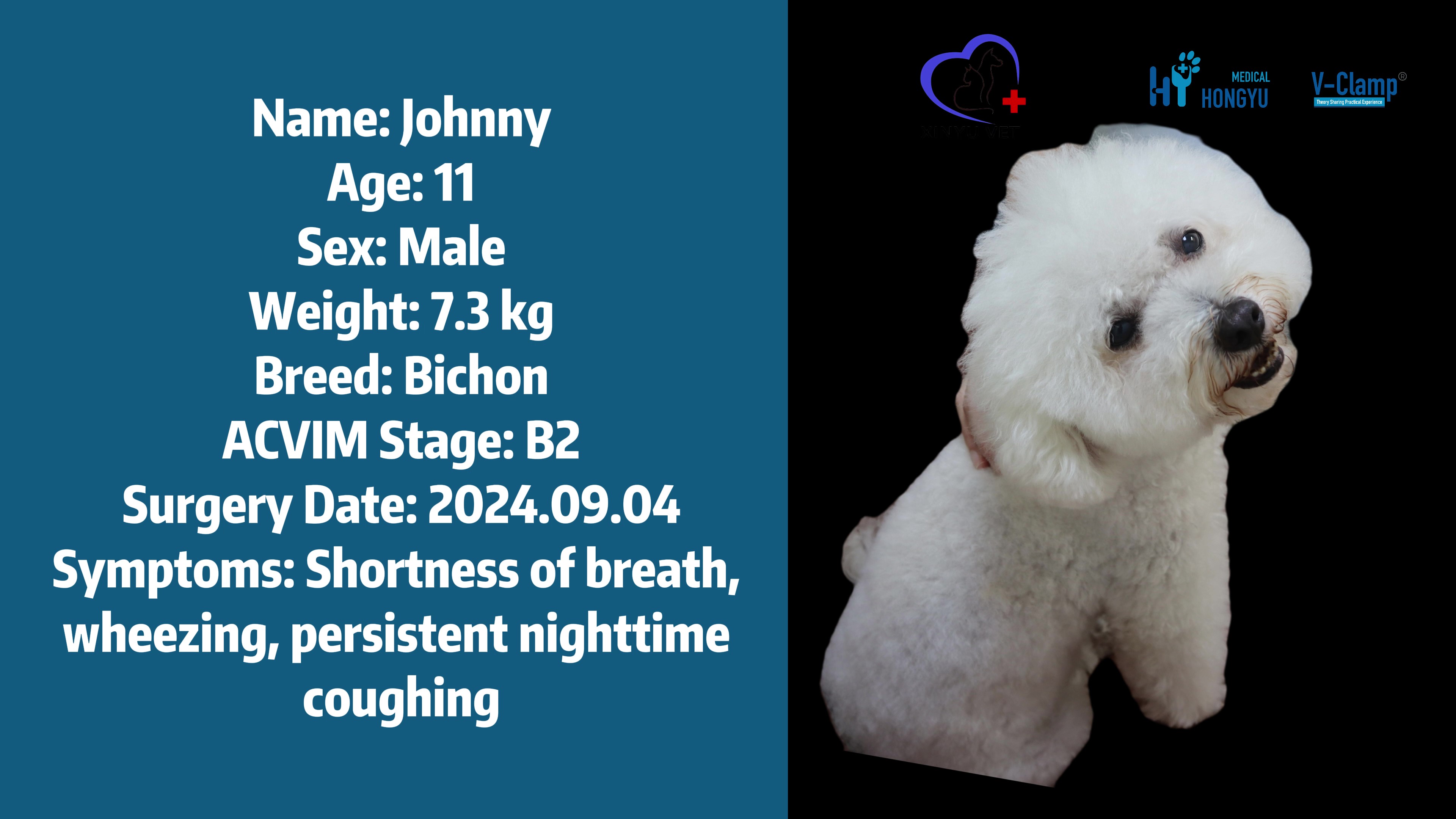
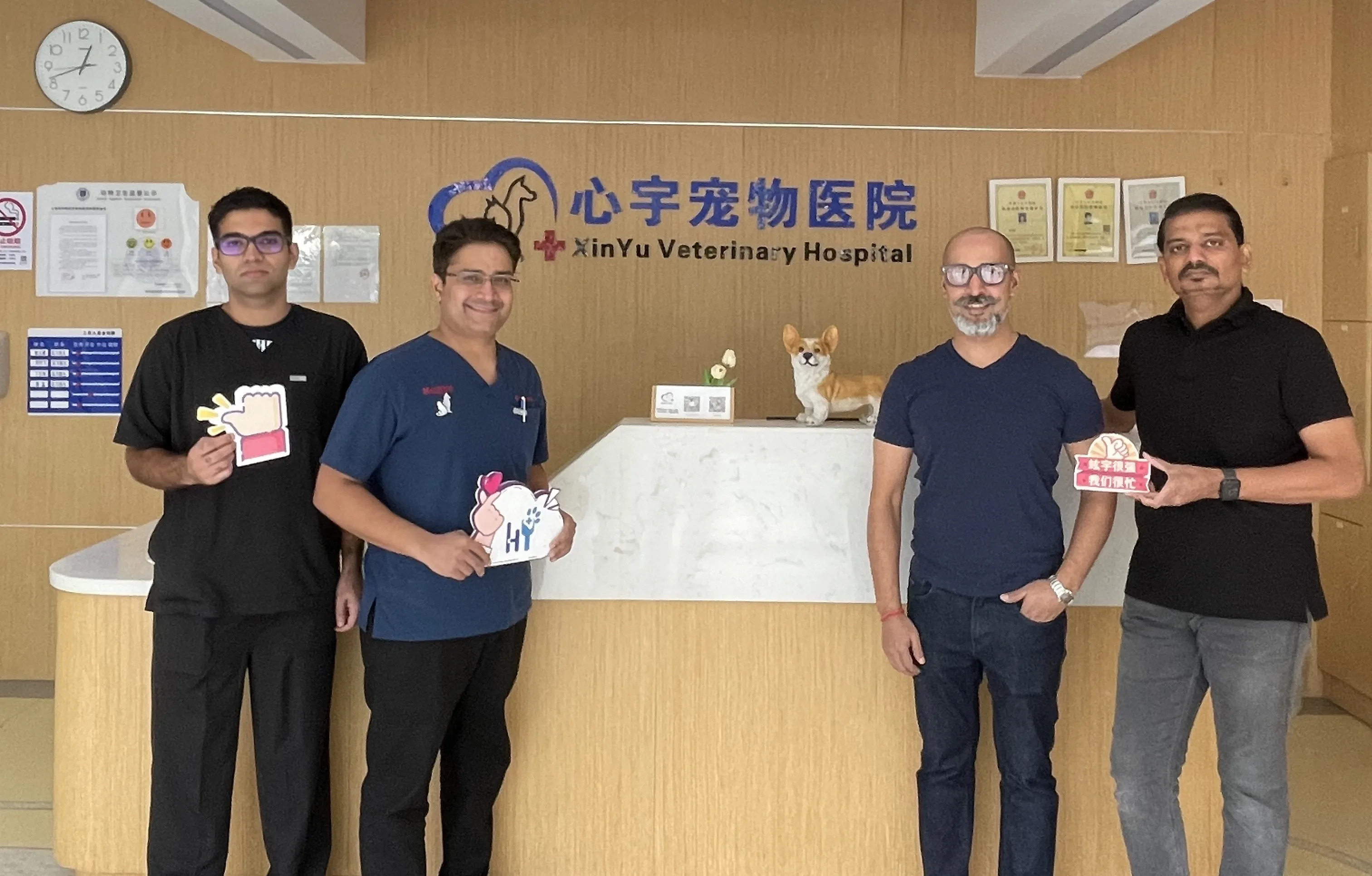
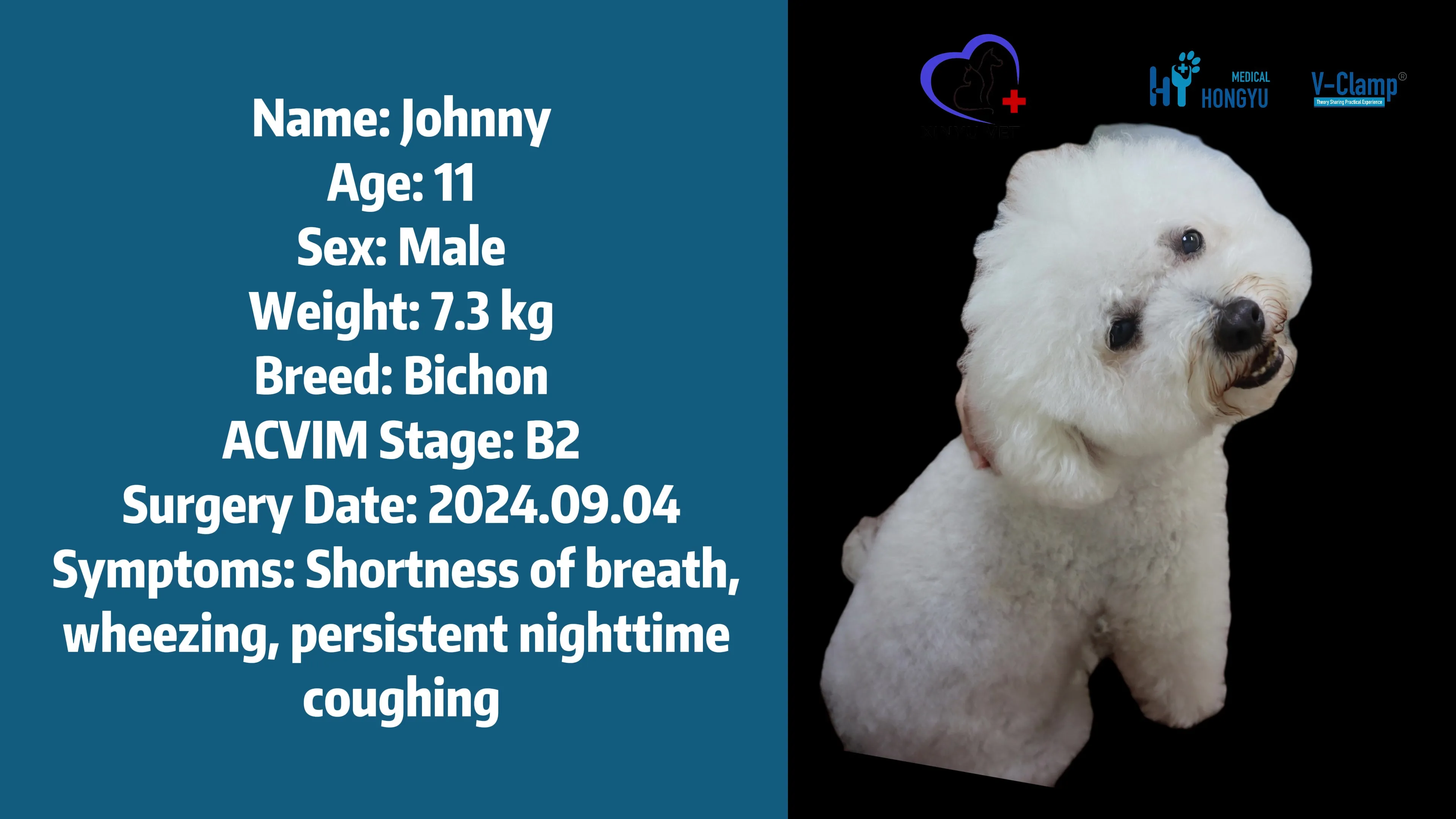
On September 4th, Shanghai Xinyu Hospital received an 11-year-old patient named Johnny, diagnosed with MMVD. A week ago, his owner noticed he suddenly had difficulty breathing, was panting, and coughing continuously during the night. Johnny's owner immediately took him to the hospital for an examination, where he was diagnosed with mitral valve regurgitation and mild tricuspid valve regurgitation. After researching online, Johnny's owner learned about Xinyu and the V-Clamp device. The day after the diagnosis, she brought Johnny to Shanghai Xinyu Pet Hospital. Xinyu’s doctors conducted a more detailed examination and confirmed that Johnny had stage B2 degenerative mitral valve disease(MMVD) with significant regurgitation. After a preliminary assessment, the doctors determined that Johnny was a candidate for the V-Clamp surgery and scheduled the operation. In the week leading up to the surgery, the doctors prescribed Johnny heart medication and diuretics to manage his condition.
On the day of the surgery, the Xinyu medical team, along with the Hongyu technical team, conducted a thorough and meticulous preoperative screening for Johnny. This included measuring his blood pressure, performing an electrocardiogram (ECG), taking X-rays, conducting blood tests, and performing a transesophageal echocardiogram (TEE).
Clinical Presentation
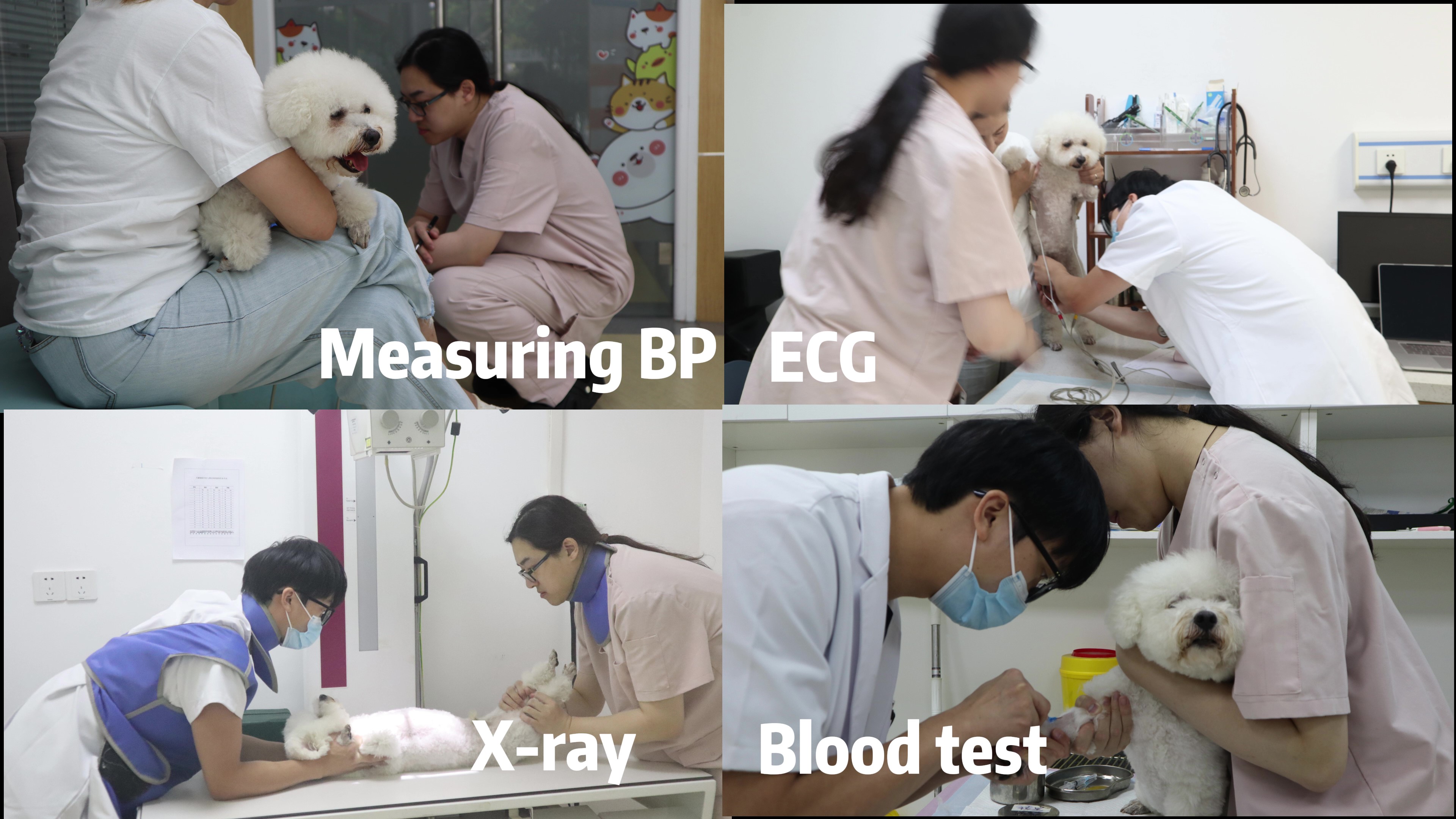
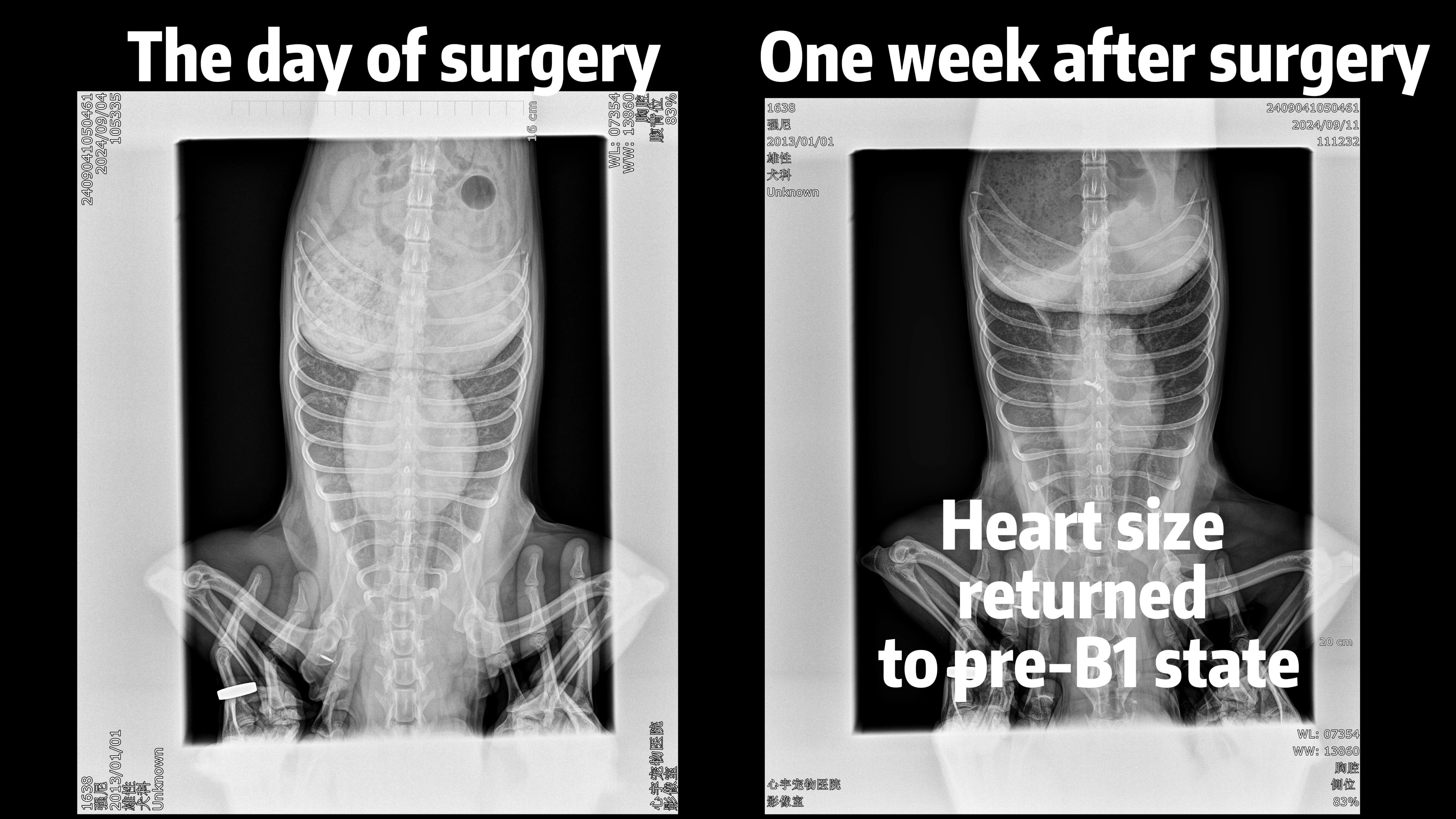
The ECG showed a normal heart rate. The X-ray revealed that Johnny’s heart had significantly enlarged compared to the time of diagnosis. Despite a week of cardiac medication and diuretic treatment, Johnny's heart continued to enlarge. A week ago, X-rays taken at another hospital showed no signs of tracheal elevation, but today's examination revealed tracheal elevation, indicating that the left atrium had enlarged and was pressing on the trachea. The blood test results showed elevated creatinine and blood urea nitrogen levels, suggesting that the diuretics had already begun to damage Johnny's kidneys.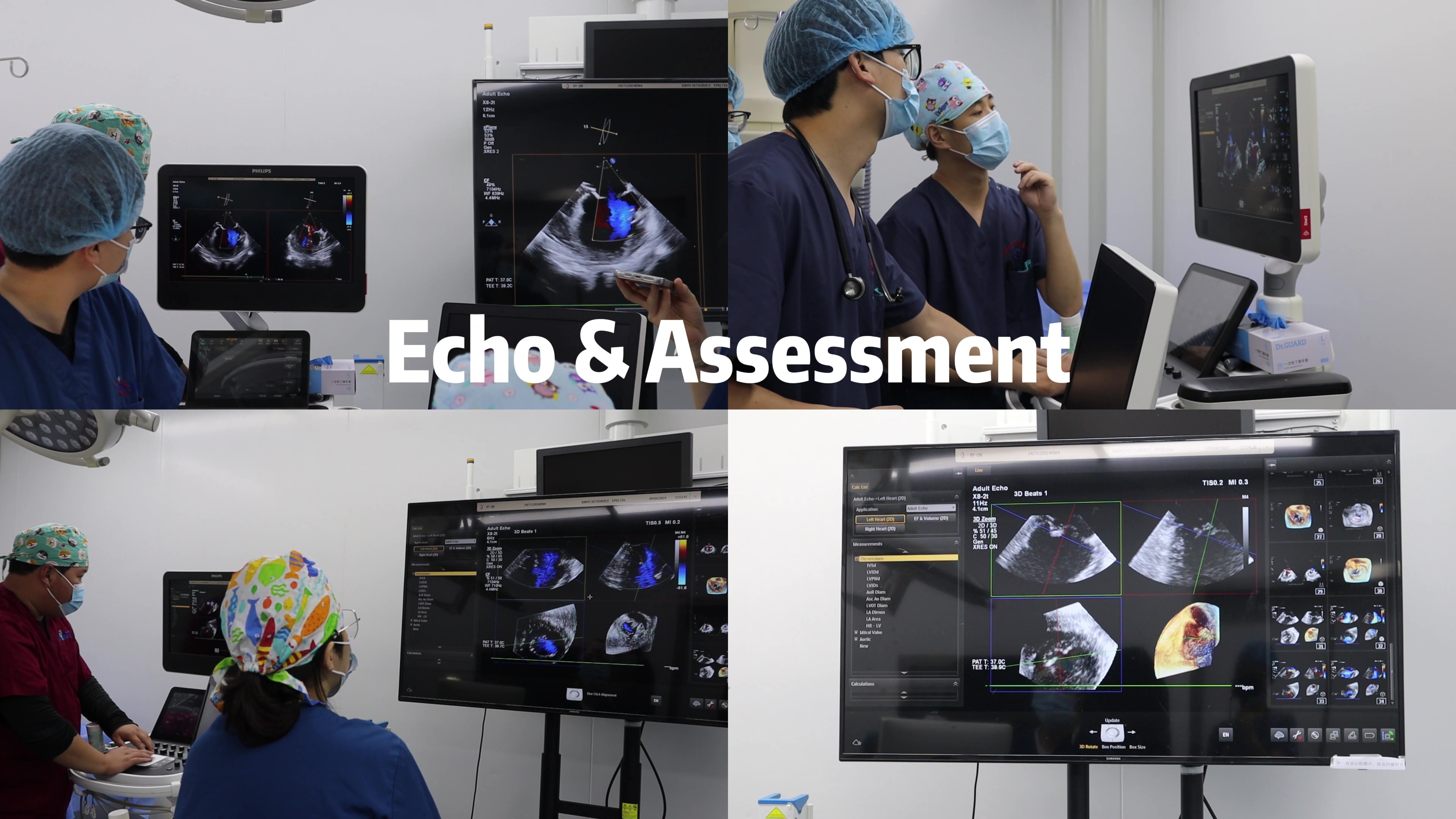
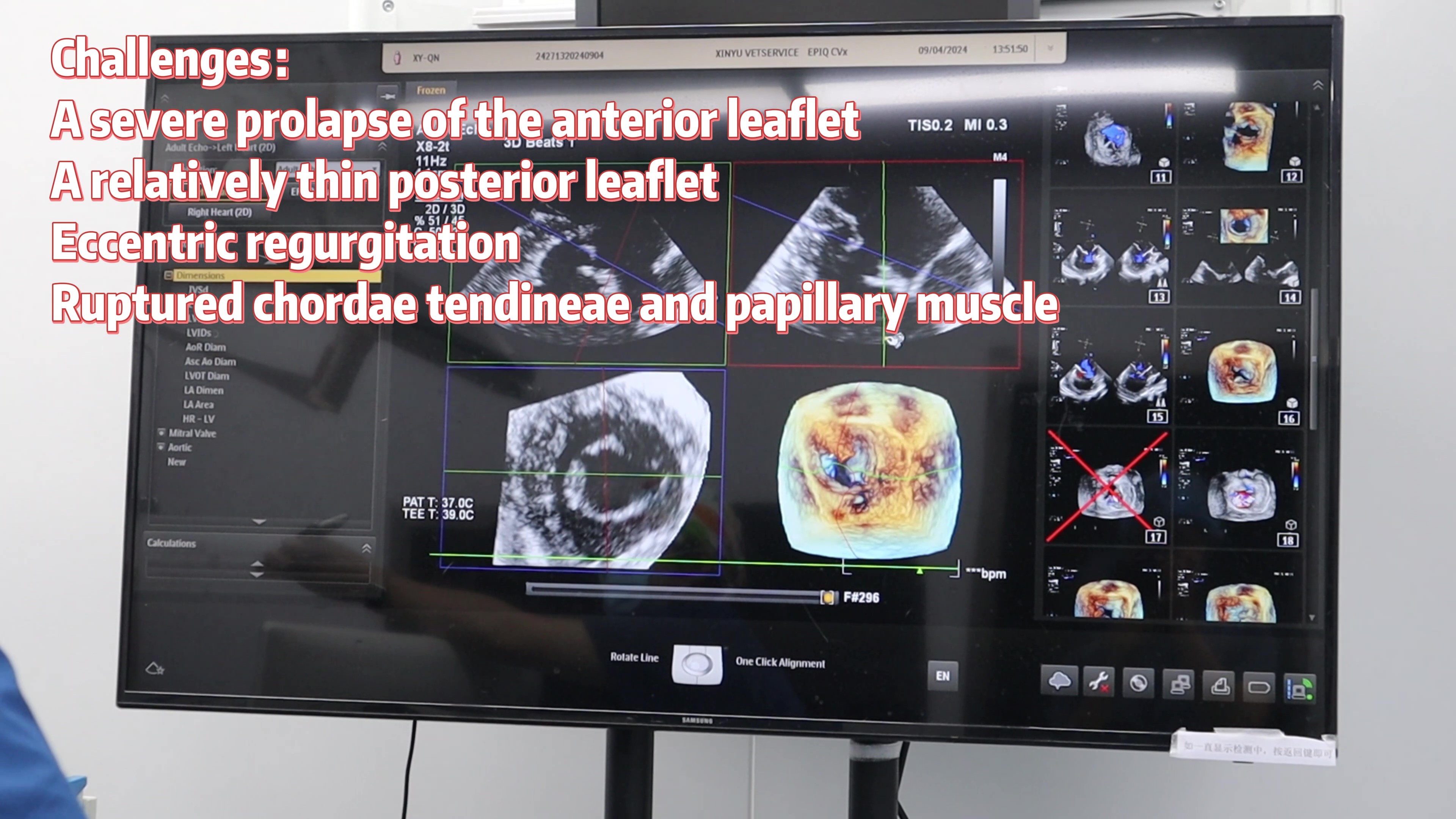
The echocardiogram showed a severe prolapse of the anterior leaflet, a relatively thin posterior leaflet (a thickness of 0.8 mm), a large regurgitation area, and an eccentric regurgitation (the coaptation line of the anterior and posterior leaflets was not perpendicular to the aorta, with a slight angle deviation).
Procedure & Outcome
After thorough analysis by the doctors and the technical team, it was confirmed that the prolapse of the anterior leaflet was due to ruptured chordae tendineae and papillary muscle.
As a result, the surgery carried significant risks and posed substantial challenges. We informed Johnny's owner of the surgical risks, explaining that, based on the cardiac assessment, even after the procedure, the improvement in regurgitation might not be as effective as initially anticipated, with only a 50%-60% improvement expected. Despite understanding all the risks, the owner decisively chose to proceed with the surgery and entrusted Johnny to our team.
Follow-up & Insights
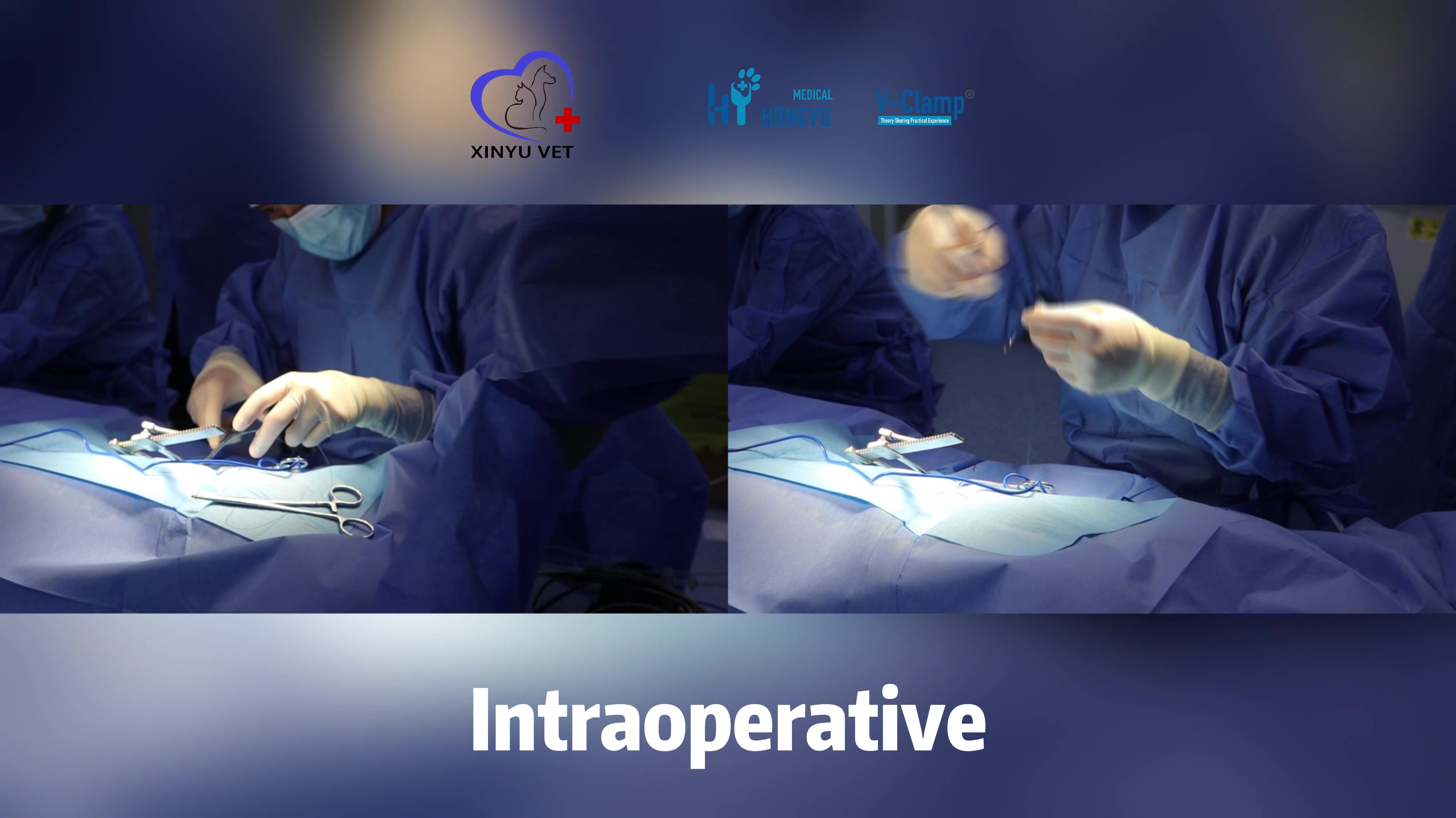
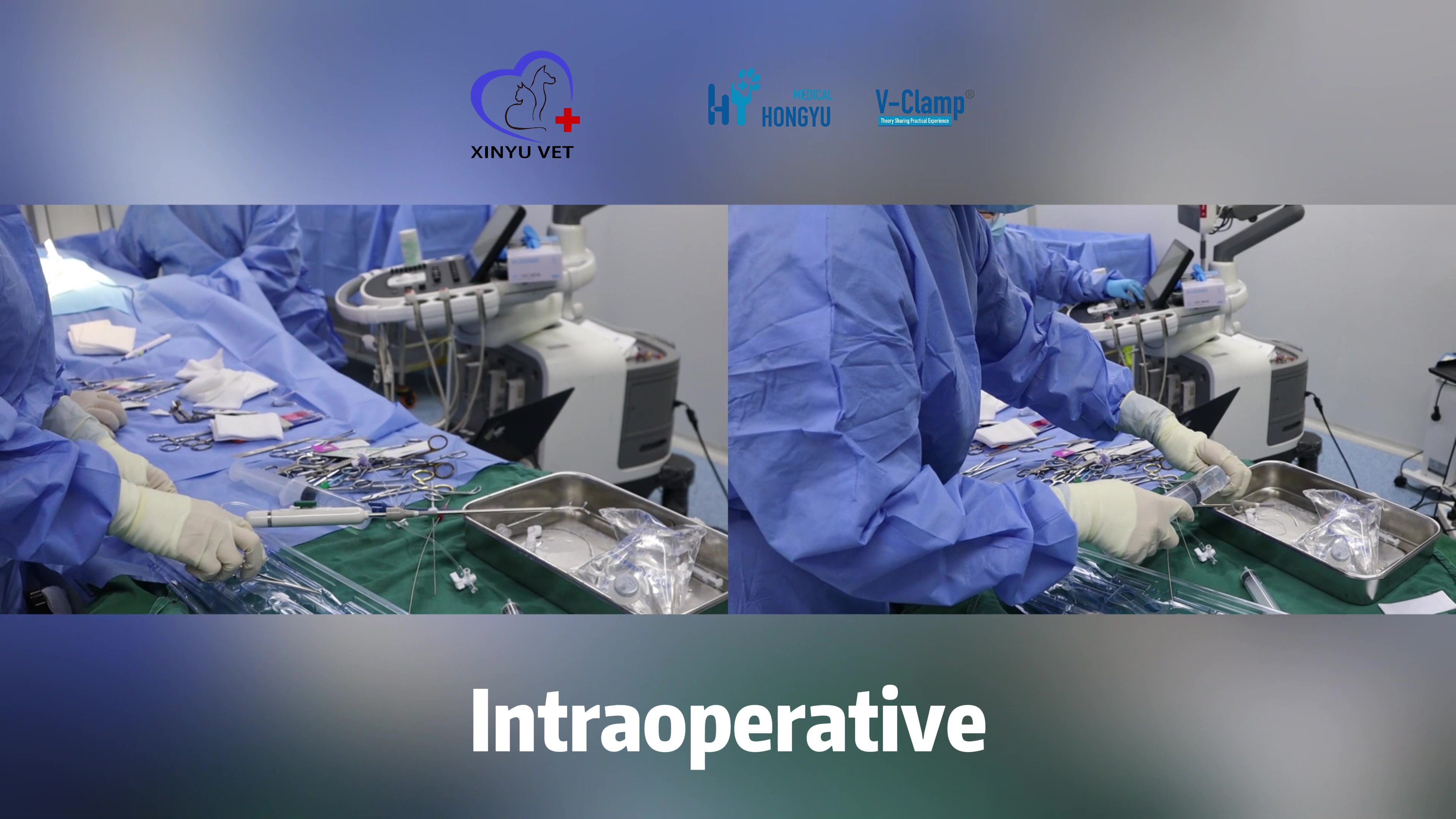
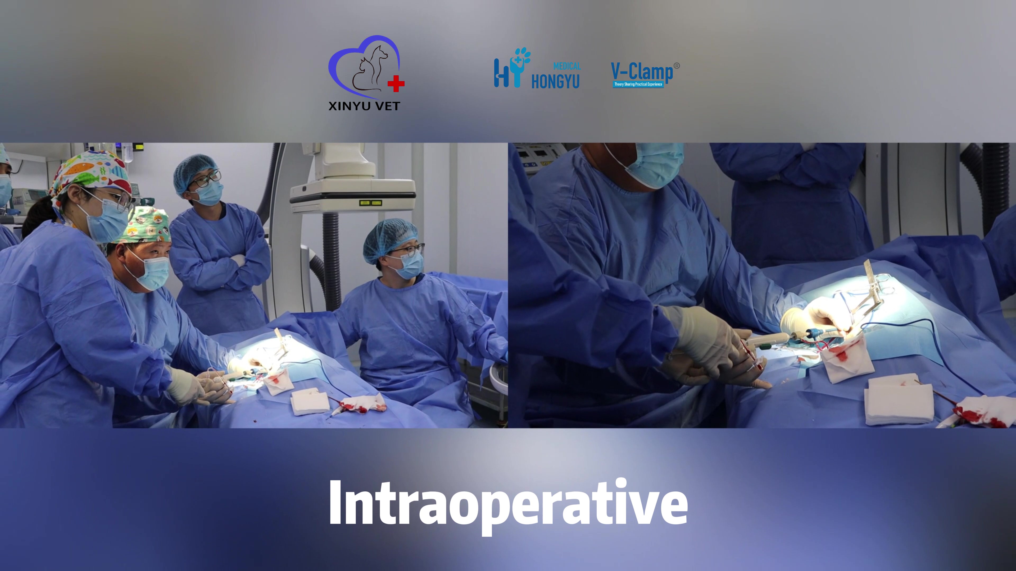
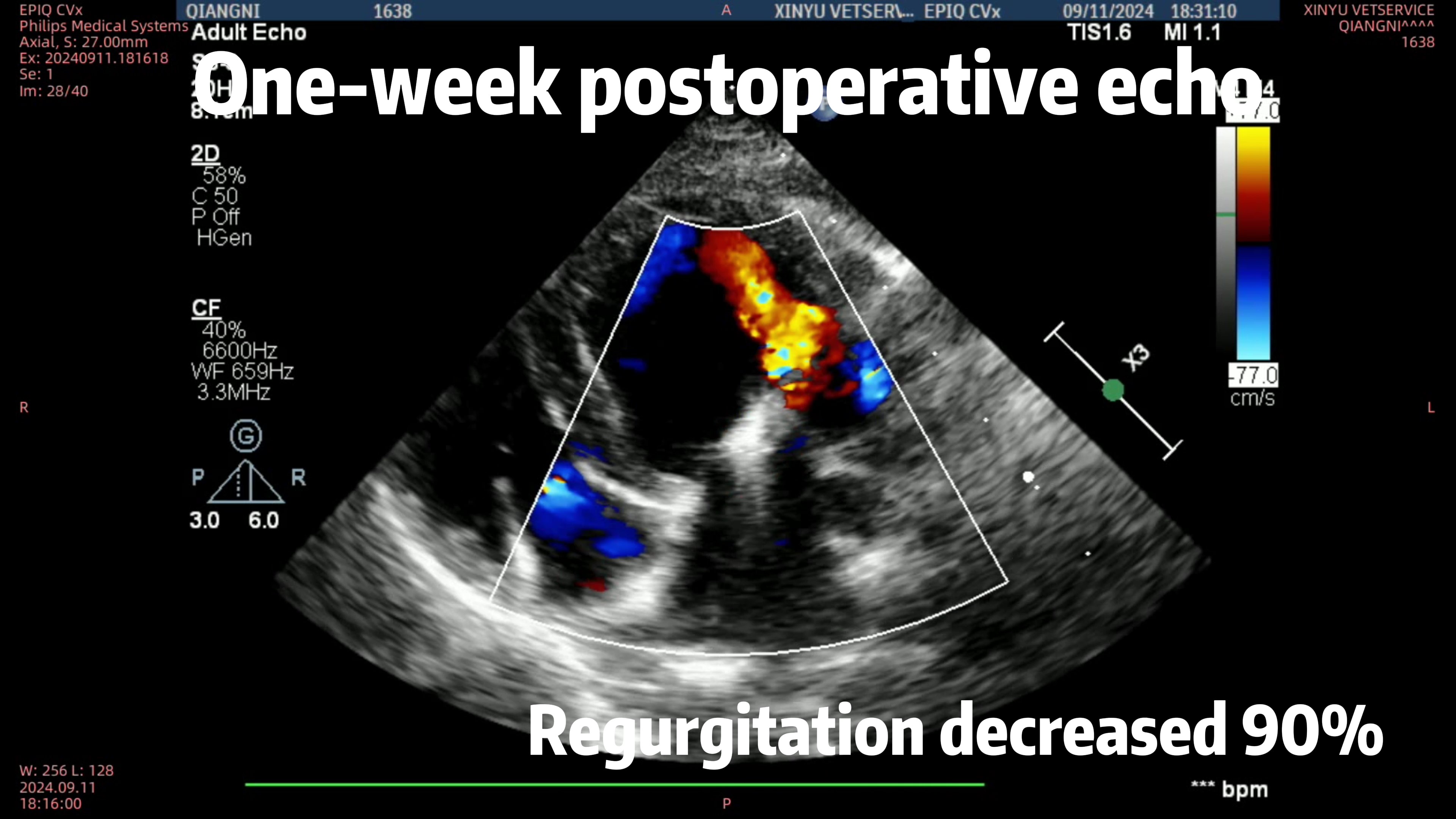
During the surgery, there was a sense of nervousness among the team, both due to the challenges and uncertainties of the procedure and the anxious expectations of Johnny’s owner. To everyone’s surprise, the surgery proceeded very smoothly. After the clipping was completed, the regurgitation was significantly reduced, with an 90% improvement, far exceeding our expectations.
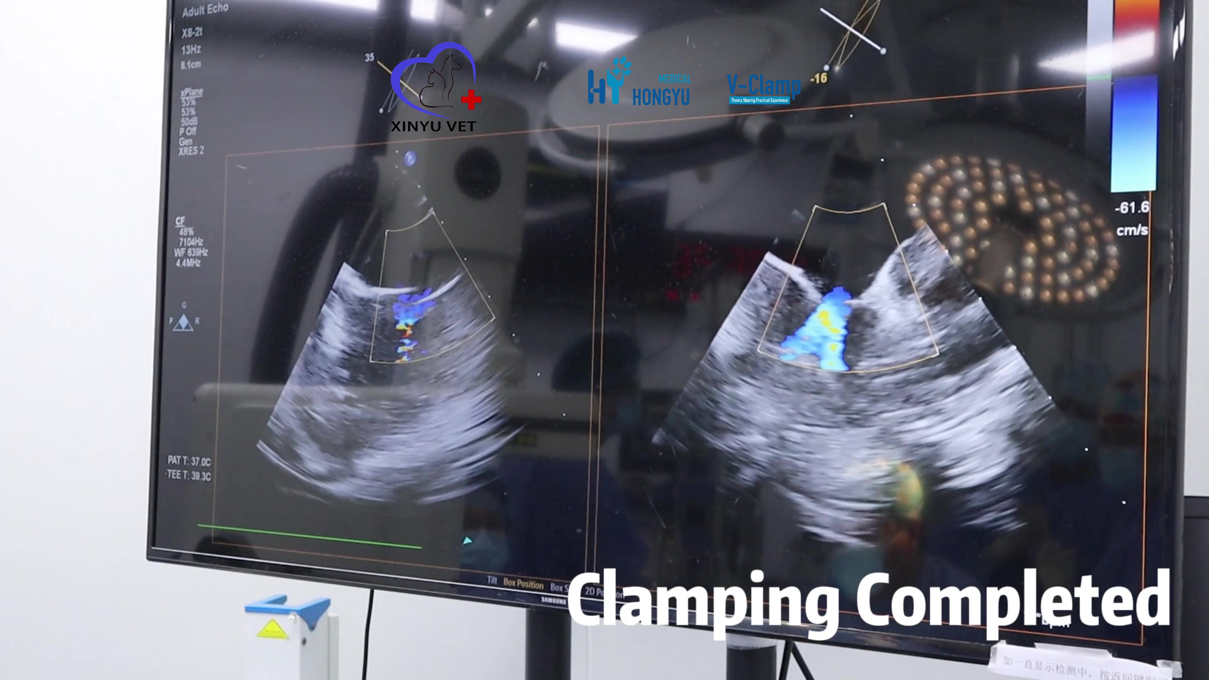
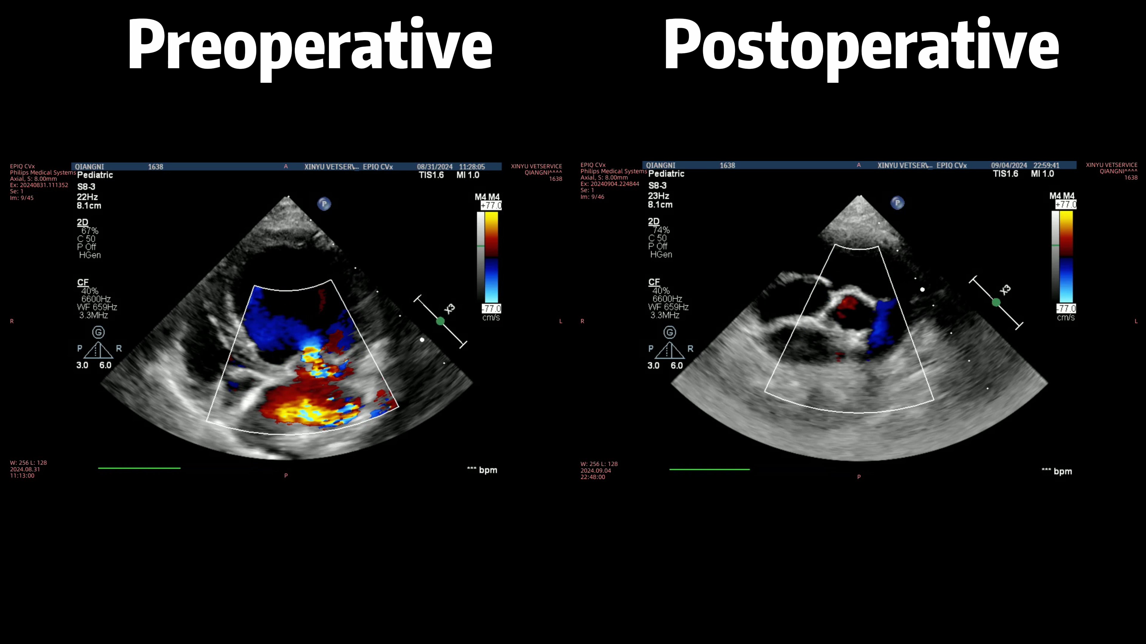
Johnny woke up quickly after the surgery, with stable anesthesia throughout the surgery and the dosage perfectly controlled. The success of the surgery was made by the close collaboration of the anesthesiologist, surgeon, cardiologist, and ultrasound specialist, as well as the trust placed in us by Johnny's owner.
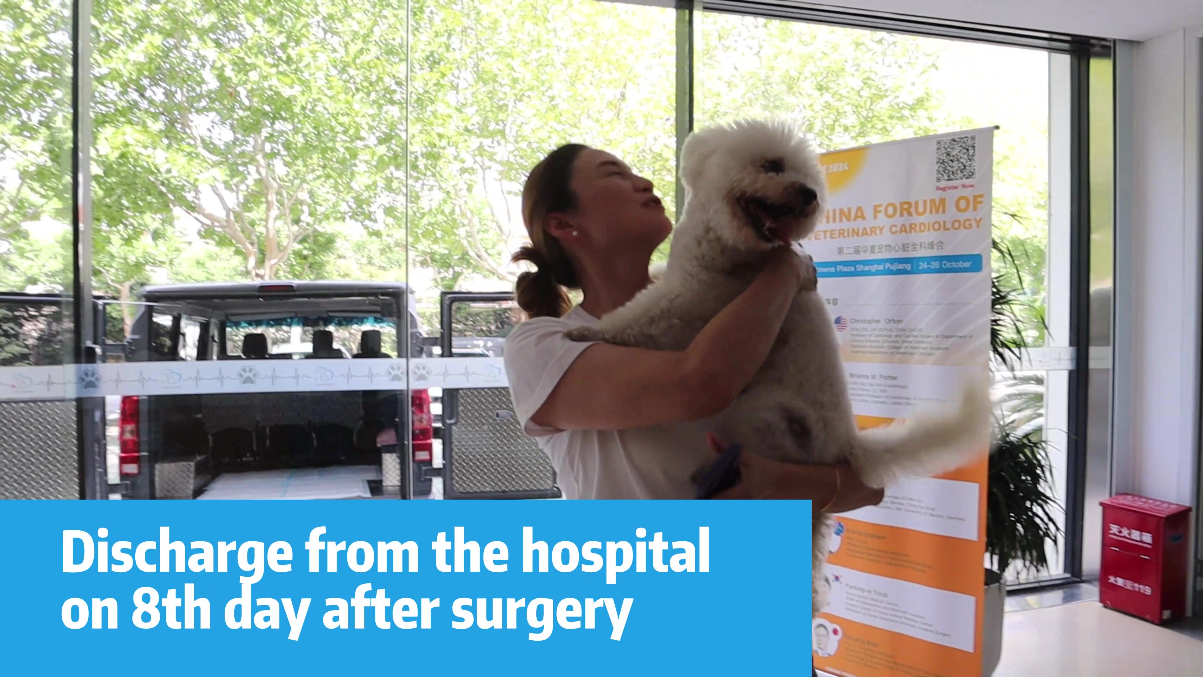
During the following week of post-operative hospitalization, Johnny's condition improved well, and he was discharged on the eighth day after surgery. The results of the check-up one week after surgery showed that all indicators had returned to normal ranges: kidney function indicators normalized, the heart size returned to its pre-B1 state, and only a small amount of regurgitation remained.
Discussion
Dr. Jing advised Johnny's owner to avoid high-intensity exercise and excessive emotional excitement for the time being. They can temporarily stop the heart medication and diuretics for a few days to observe, but Johnny will need to take anticoagulants for 30 days (once a day) and antibiotics for 3 days (twice a day), and return for a follow-up check in two weeks.
Closing Notes
The success of this surgery can largely be attributed to Johnny’s owner, who promptly recognized the dog's condition, sought medical evaluation and treatment, and placed unwavering trust in Xinyu, making the decisive choice to proceed with surgery. The timing of the procedure was critical. Given Johnny’s larger size, if the condition had continued to progress, the annulus AP diameter could have exceeded 20 mm, rendering the V-Clamp surgery unsuitable.
The quality of the valve leaflets also plays a key role in the success of the V-Clamp procedure. Had the surgery been delayed by another 1-2 months, the progression of MMVD would have further weakened the valve leaflets due to regurgitation flush, making surgery more difficult. While many cases undergo surgery at stage C or even D, Johnny’s operation at stage B2 provided a better opportunity for recovery, as the heart had not yet undergone significant remodeling and degeneration, and the valve leaflets remained in better condition.